. 2011 Oct;31(19):3963-74.
doi: 10.1128/MCB.05297-11. Epub 2011 Aug 1.

Phosphotyrosine-dependent coupling of Tim-3 to T-cell receptor signaling pathways

Affiliations

Phosphotyrosine-dependent coupling of Tim-3 to T-cell receptor signaling pathways

Judong Lee et al. Mol Cell Biol. 2011 Oct.

Abstract

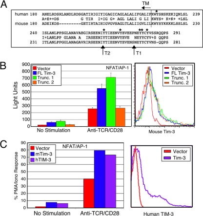
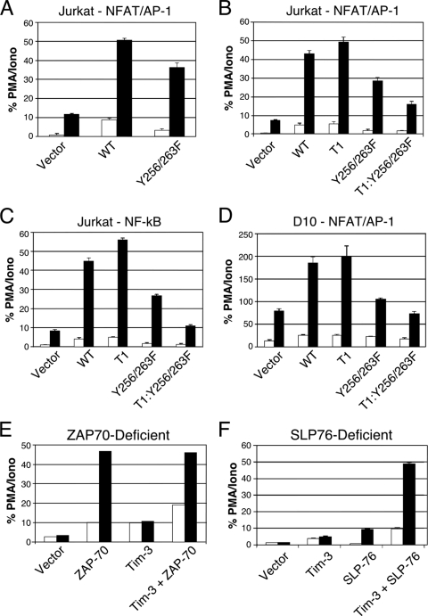
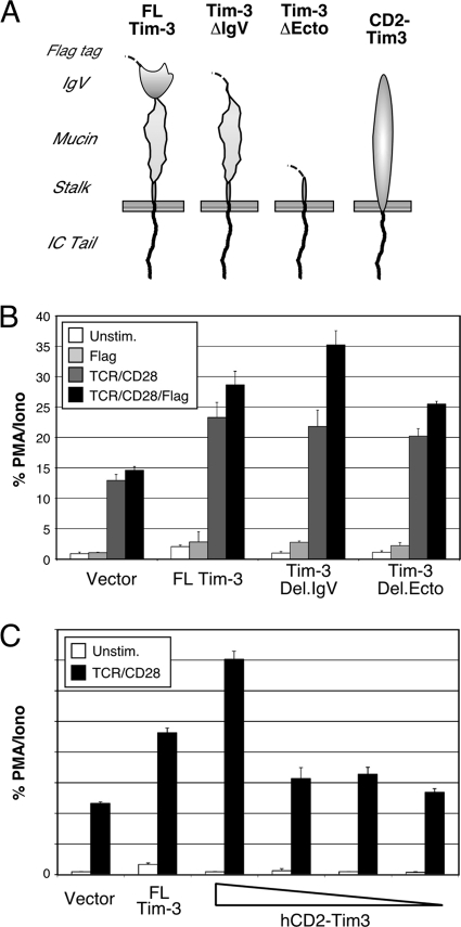
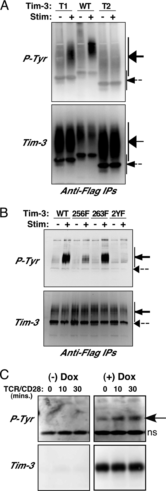
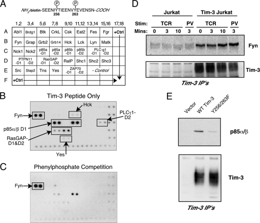
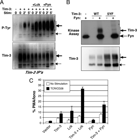
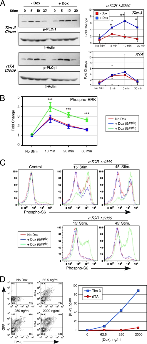
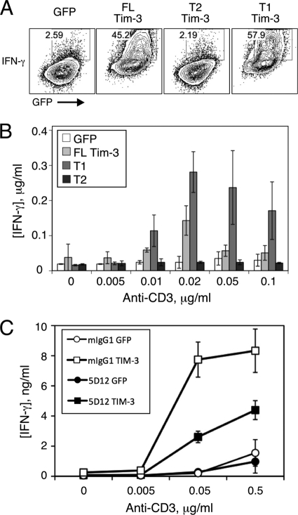
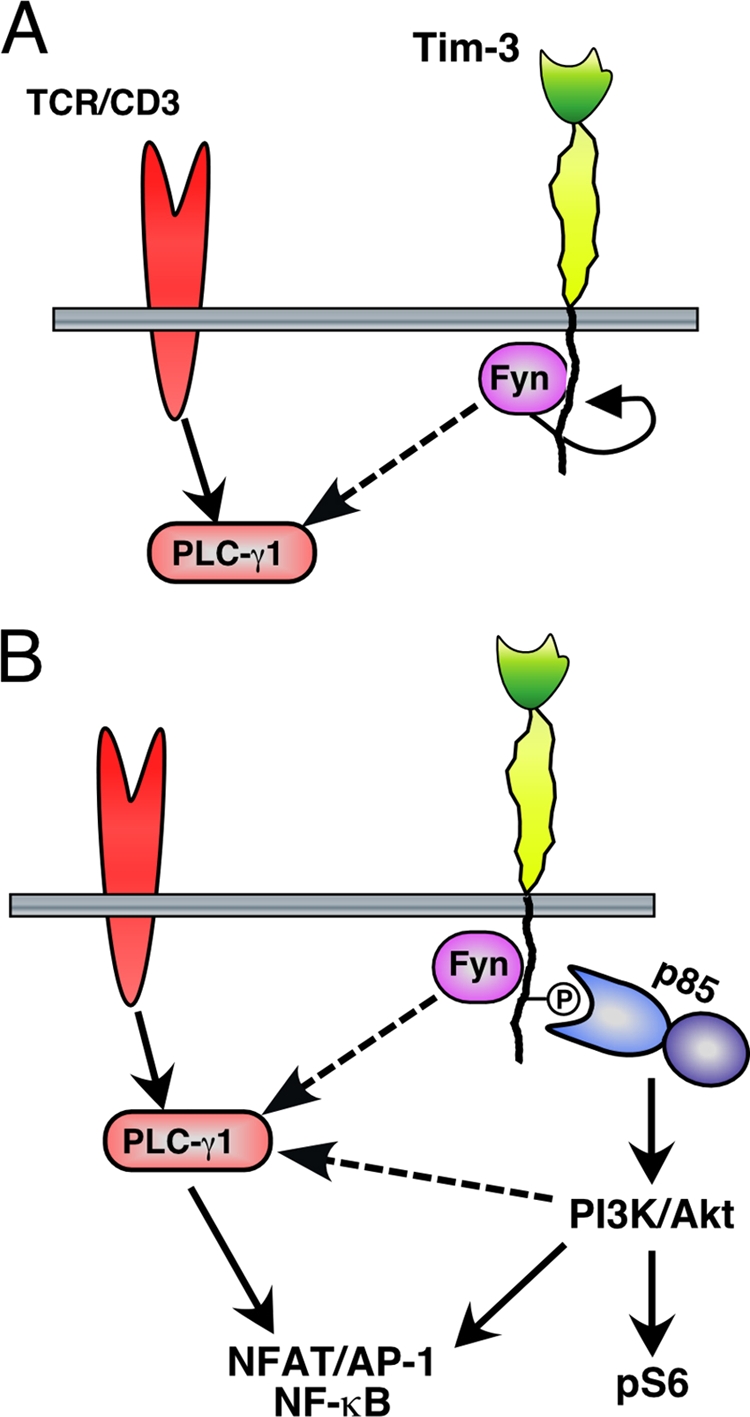
The transmembrane protein Tim-3 has been shown to negatively regulate T-cell-dependent immune responses and was recently demonstrated to be associated with the phenomenon of immune exhaustion, which can occur as a consequence of chronic viral infection. Unlike other negative regulators of T-cell function (e.g., PD-1), Tim-3 does not contain any obvious inhibitory signaling motifs. We have found that ectopic expression of Tim-3 in T cells leads to enhancement of T-cell receptor (TCR)-dependent signaling pathways, which was observed at the level of transcriptional reporters and endogenous cytokine production. We have exploited this observation to dissect what elements within the cytoplasmic tail of Tim-3 are required for coupling to downstream signaling pathways. Here we have demonstrated that two of the more membrane-proximal cytoplasmic tail tyrosines are required for Tim-3 signaling to T-cell activation pathways in a redundant fashion. Furthermore, we show that Tim-3 can directly bind to the Src family tyrosine kinase Fyn and the p85 phosphatidylinositol 3-kinase (PI3K) adaptor. Thus, at least under conditions of short-term stimulation, Tim-3 can augment T-cell activation, although this effect can be blocked by the inclusion of an agonistic antibody to Tim-3. These findings should help further the study of Tim-3 function in other physiological settings, such as those that lead to immune exhaustion.


Figures

Fig. 1.
Fig. 1.
Ectopic Tim-3 expression augments activation of NFAT/AP-1 and NF-κB. (A) Alignment of the C-terminal regions of murine and human Tim-3. The boundary of the TM/cytoplasmic tail is indicated, as are truncations discussed below. Relevant tyrosines are indicated with asterisks. (B) Effect of ectopic wild-type or truncated (Trunc.) Tim-3 expression on NFAT/AP-1 induction in Jurkat T cells (left panel). Luciferase activity is presented as absolute light units. Equivalent transfection efficiency was confirmed by expression of a cotransfected GFP (not shown). Equal expression of the constructs was confirmed by flow cytometry (right panel). (C) Enhancement of NFAT/AP-1 induction by human TIM-3. Jurkat T cells were transfected with Flag-tagged mTim-3 or untagged hTIM-3. NFAT/AP-1 luciferase activity was determined after a 6-h stimulation (left panel). Human TIM-3 expression was confirmed by flow cytometry (right panel). Luciferase results in panel C are the percentage of the maximal stimulation obtained with PMA-ionomycin (PMA/Iono). Results shown in panels B and C are the average ± SD for triplicate samples from a single experiment, representative of at least three that were performed in each case.
Fig. 2.
Fig. 2.
Requirements for transcriptional activation by Tim-3. Jurkat T cells (A to C) were transfected with an NFAT/AP-1 (A and B) or NF-κB (C) reporter plus the indicated Tim-3 constructs. D10 T cells (D) were also transfected with an NFAT/AP-1 reporter and the indicated Tim-3 constructs. Jurkat T-cell lines lacking expression of either ZAP-70 (E) or SLP-76 (F) were transfected with NFAT/AP-1-luciferase and the indicated constructs. The next day, cells were left unstimulated (white bars) or stimulated with anti-TCR/CD28 monoclonal antibodies (MAbs) (black bars). Luciferase activity is presented as the percentage of maximal stimulation obtained with PMA-ionomycin. Luciferase activity is presented as the percentage of the maximal response obtained with PMA plus ionomycin. Results shown are the average ± SD of data for triplicate samples from a single experiment, representative of at least three that were performed in each case.
Fig. 3.
Fig. 3.
The cytoplasmic domain of Tim-3 is sufficient to costimulate TCR/CD28-induced NFAT/AP-1. Jurkat T cells were transfected with an NFAT/AP-1 reporter and the indicated constructs and then stimulated as indicated before determination of luciferase activity. (A) Schematic of Tim-3 constructs used in these experiments. (B) Effect of ectodomain deletions on Tim-3 costimulation of NFAT/AP-1. (C) Costimulation of NFAT/AP-1 activation by a CD2-Tim3 chimera.
Fig. 4.
Fig. 4.
Availability of Tim-3 cytoplasmic tyrosine residues for phosphorylation. (A and B) 293T cells were transfected with the indicated Flag-tagged Tim-3 constructs and then split and left unstimulated or treated with pervanadate. Lysates were prepared and subjected to IP with an anti-Flag antibody, followed by SDS-PAGE and Western blotting. Blots were first probed for Tim-3 (lower panel) and then stripped and reprobed with an antiphosphotyrosine antibody (upper panel). (C) Parental Jurkat T cells or Jurkat T cells stably transfected with Flag-tagged Tim-3 were stimulated as indicated and prepared for IP and Western blotting as described above. After IP, samples were treated with PNGase F prior to SDS-PAGE.
Fig. 5.
Fig. 5.
Identification of SH2 domain-containing proteins capable of interacting with the cytoplasmic tail of Tim-3. (A) Detail of the phospho-peptide derived from the Tim-3 sequence (top) used to screen a solid-phase array containing recombinant SH2 domains from the indicated proteins (bottom). (B) Representative results of screening the array with the Tim-3 phospho-peptide. (C) Representative results of a screen performed with the same array and peptide, except in the presence of 200 mM phenyl phosphate. (D) Confirmation of activation-independent interaction of Fyn with Tim-3. Control or Tim3-expressing Jurkat T cells were stimulated (Stim) for the indicated times with anti-TCR antibody (TCR) or pervanadate (PV), lysed, and subjected to IP with anti-Flag antibody. Western blots were probed for endogenous Fyn (upper panel) or the transfected Tim-3 (lower panel). (E) Confirmation of p85 PI3K binding to Tim-3. Jurkat T cells were transfected with the indicated plasmids and stimulated with pervanadate. Anti-Flag IPs were performed and analyzed by blotting for p85, followed by anti-Flag.
Fig. 6.
Fig. 6.
Role of Src kinases in Tim-3 phosphorylation and function. (A) 293T cells were transfected with full-length Tim-3, alone or with Lck or Fyn. Cells were stimulated with pervanadate for the indicated times. Tim-3 was IP'd from lysates, and the presence of Tim-3 was first confirmed by Western blotting (lower panel), followed by stripping and reprobing with an antiphosphotyrosine (P-Tyr) antibody (upper panel). (B) In vitro kinase assay showing direct phosphorylation of Tim-3 by Fyn (upper panel). Purified wild-type Tim-3 or a version lacking all cytoplasmic tyrosines (except for the membrane proximal tyrosine) was subjected to an in vitro kinase assay with purified active Fyn kinase. Levels of purified Tim-3 were confirmed by Western blotting (lower pnael). (C) Lck-deficient JCaM.1 cells were transfected with an NFAT/AP-1 reporter and the indicated plasmids. Cells were stimulated the next day as indicated. Luciferase activity is presented as the percentage of the maximal response obtained with PMA and ionomycin. Tim-3 expression was verified by flow cytometry (not shown).
Fig. 7.
Fig. 7.
Augmented TCR signaling and cytokine production by Tim-3. (A) Tet-on-Tim3 or parental Jurkat T cells expressing only the reverse tetracycline transactivator (rtTA) were cultured for 2 days without or with doxycycline (Dox). Cells were stimulated as indicated for analysis of phospho-PLC-γ1 (Y783) by Western blotting, followed by probing for beta-actin as a loading control (left). Average results (± SD) from five separate experiments are shown (*, P < 0.05; **, P < 0.01) (right). (B and C) Tet-on-Tim3 Jurkat T cells were cultured for 2 days without or with Dox. Cells were then stimulated as indicated and processed for intracellular staining of phospho-ERK (average of results from three experiments ± SD; ***, P < 0.001) (B) or phospho-S6 (C). In addition, Dox-treated cells were subdivided into GFPlo (nonexpressing) and GFPhi (Tim3 expressing). (D) Tet-on-Tim3 Jurkat T cells were cultured with the indicated concentrations of Dox. Analysis by flow cytometry of Tim-3 expression (x axis) and a coexpressed GFP reporter (y axis) is shown (left). Cells were stimulated for 24 h with soluble anti-TCR/CD28 antibodies; cell-free supernatants were harvested, and secreted IL-2 was measured by ELISA (right).
Fig. 8.
Fig. 8.
Costimulation of acute T-cell cytokine production by ectopic Tim-3 expression. Naive murine CD4+ T cells were stimulated with anti-CD3/CD28 antibodies under Th1 conditions and then infected with retrovirus encoding the indicated constructs and restimulated with the indicated concentrations of soluble anti-CD3 antibody, again under Th1 conditions. Two days later, cells were processed for intracellular cytokine staining and flow cytometry. (A) Representative flow cytometry of intracellular IFN-γ and retroviral coexpressed GFP. (B and C) T cells were restimulated with the indicated concentrations of anti-CD3, and supernatant was collected for determination of IFN-γ concentrations by ELISA.
Fig. 9.
Fig. 9.
Model for acute enhancement of TCR signaling and T-cell activation by Tim-3.

References

    1. Anderson A. C., et al. 2007. Promotion of tissue inflammation by the immune receptor Tim-3 expressed on innate immune cells. Science 318:1141–1143 - PubMed
    1. Angelosanto J. M., Wherry E. J. 2010. Transcription factor regulation of CD8+ T-cell memory and exhaustion. Immunol. Rev. 236:167–175 - PubMed
    1. Apetoh L., et al. 2010. The aryl hydrocarbon receptor interacts with c-Maf to promote the differentiation of type 1 regulatory T cells induced by IL-27. Nat. Immunol. 11:854–861 - PMC - PubMed
    1. Barber D. L., et al. 2006. Restoring function in exhausted CD8 T cells during chronic viral infection. Nature 439:682–687 - PubMed
    1. Binne L. L., Scott M. L., Rennert P. D. 2007. Human TIM-1 associates with the TCR complex and up-regulates T cell activation signals. J. Immunol. 178:4342–4350 - PubMed

Publication types

MeSH terms