. 2011 Oct;31(19):3924-37.
doi: 10.1128/MCB.01269-10. Epub 2011 Aug 1.

RSC facilitates Rad59-dependent homologous recombination between sister chromatids by promoting cohesin loading at DNA double-strand breaks

Affiliations

RSC facilitates Rad59-dependent homologous recombination between sister chromatids by promoting cohesin loading at DNA double-strand breaks

Ji-Hyun Oum et al. Mol Cell Biol. 2011 Oct.

Abstract

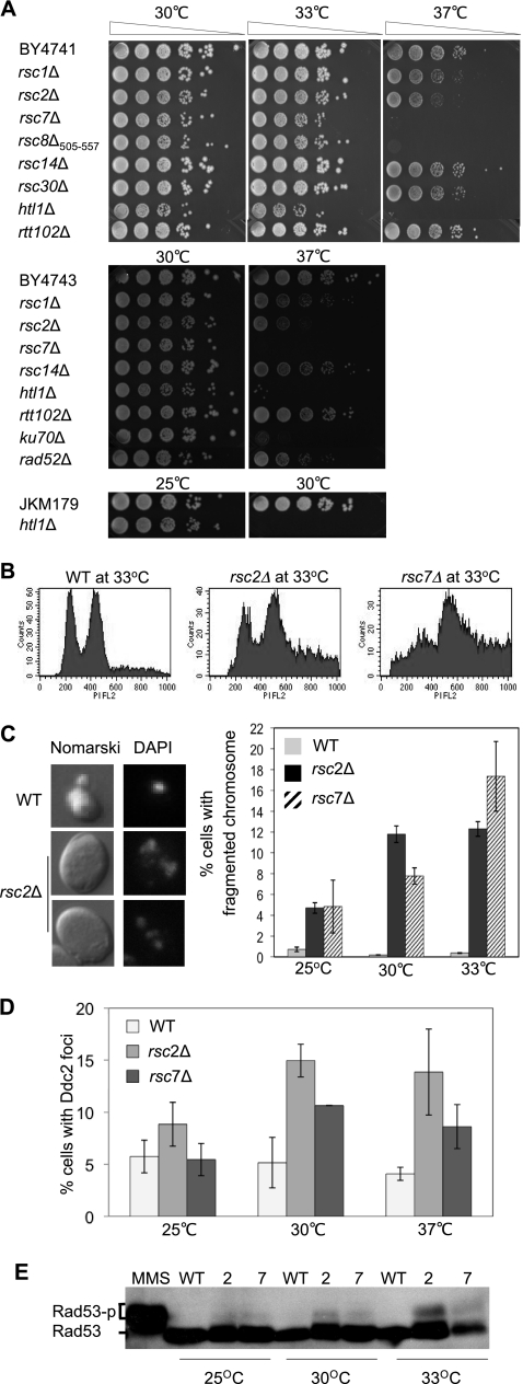
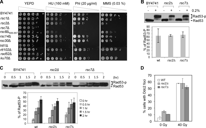
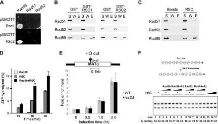
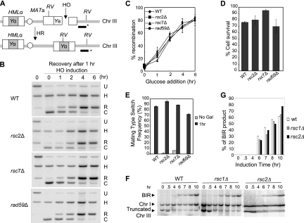
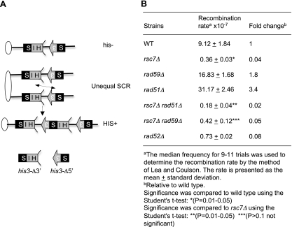
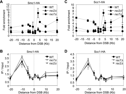
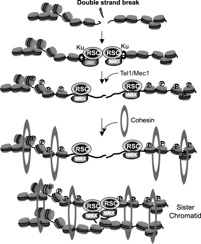
Homologous recombination repairs DNA double-strand breaks by searching for, invading, and copying information from a homologous template, typically the homologous chromosome or sister chromatid. Tight wrapping of DNA around histone octamers, however, impedes access of repair proteins to DNA damage. To facilitate DNA repair, modifications of histones and energy-dependent remodeling of chromatin are required, but the precise mechanisms by which chromatin modification and remodeling enzymes contribute to homologous DNA repair are unknown. Here we have systematically assessed the role of budding yeast RSC (remodel structure of chromatin), an abundant, ATP-dependent chromatin-remodeling complex, in the cellular response to spontaneous and induced DNA damage. RSC physically interacts with the recombination protein Rad59 and functions in homologous recombination. Multiple recombination assays revealed that RSC is uniquely required for recombination between sister chromatids by virtue of its ability to recruit cohesin at DNA breaks and thereby promoting sister chromatid cohesion. This study provides molecular insights into how chromatin remodeling contributes to DNA repair and maintenance of chromatin fidelity in the face of DNA damage.


Figures

Fig. 1.
Fig. 1.
rsc mutations induce a spontaneous DNA damage response. (A) Mutants lacking nonessential RSC subunits in the BY4741, BY4743 diploid, and JKM179 backgrounds were tested for growth at 30°C, 33°C, and 37°C. Fivefold serial dilutions were plated and photographed after 3 to 4 days of growth. (B) Cell cycle distribution was assessed by flow cytometry as described in Materials and Methods. Wild-type and rsc2 and rsc7 mutant cells grown overnight in YEPD at 33°C were analyzed by FACS. (C) The nuclear morphology of BY4741 and its rsc mutant derivatives grown at 33°C was examined by DeltaVision fluorescence microscopy (left). The images shown were obtained with Nomarski optics after 4′,6-diamidino-2-phenylindole (DAPI) staining. Percentages of cells containing fragmented nuclei were scored from at least 300 cells in each mutant and plotted as a function of increasing growth temperature (right). The results are the average of at least three independent experiments ± the standard deviation. (D) Percentages of wild-type (WT), rsc2Δ mutant, and rsc7Δ mutant cells showing Ddc2-GFP foci at the indicated growth temperatures. More than 300 cells were analyzed in each case. (E) Phosphorylation of Rad53 in wild-type cells and the rsc2 and rsc7 mutants at different growth temperatures was analyzed by Western blotting using anti-Rad53 antibody. For comparison, hyperphosphorylation of Rad53 was induced by treating logarithmically growing wild-type cells with 0.2% MMS for 20 min (lane 1).
Fig. 2.
Fig. 2.
Integrity of cell cycle checkpoint activation in rsc mutants. (A) Viability of mutants after DNA-damaging treatment was determined by spotting cells (5-fold serial dilutions) onto YEPD plates containing hydroxyurea (HU), phleomycin (Phl), or MMS. The plates were photographed after 3 to 4 days of growth at 30°C. (B) The integrity of damage-induced cell cycle checkpoint activation was assessed in cells treated with 0.2% MMS for 30 min and examined for the level of Rad53 phosphorylation by Western blotting using anti-Rad53 antibody. Percent Rad53 phosphorylation was calculated as intensity of phosphorylated Rad53/intensity of total Rad53 using the ImageJ program. The averages of three independent experiments ± the standard deviations are shown. (C) Kinetics of Rad53 phosphorylation in cells that were arrested at S/G2 and exposed to 0.033% MMS for the indicated times. Percent Rad53 phosphorylation was calculated as described for panel B. The averages of three independent experiments ± the standard deviations are shown. (D) Yeast cells expressing Ddc2-GFP were gamma irradiated and fixed with paraformaldehyde, and the number of cells showing Ddc2-GFP foci was scored using a DeltaVision microscope. Over 300 cells of each strain were analyzed. wt, wild type.
Fig. 3.
Fig. 3.
RSC participates in Rad59-dependent DNA repair. (A) The viability of single and double rsc mutant combinations and the indicated HR genes was determined by spotting cells onto culture plates containing the indicated genotoxic chemicals. Fivefold serial dilutions were spotted, and the plates were photographed after 3 to 4 days of growth at 30°C. (B) Percent survival after gamma irradiation or MMS treatment. Logarithmic-phase cultures of yeast cells were harvested, resuspended in phosphate-buffered saline, irradiated with the indicated dose of gamma rays or treated with the indicated concentration of MMS for 20 min, and plated onto YEPD plates. After 3 to 4 days, surviving colonies were counted and the survival rate was calculated by comparison to the number of colonies of mock-treated cells. Each experimental point represents the average of three independent experiments ± the standard deviation. HU, hydroxyurea.
Fig. 4.
Fig. 4.
Rad59 interacts with and enhances the ATPase activity of the RSC complex. (A) Interactions of Rsc1 and Rsc2 with Rad51, Rad52, and Rad59 were determined by yeast two-hybrid assays. Interactions were discerned based on expression of the HIS3 reporter gene by spotting cells on SC in the absence of histidine. (B) Pulldown experiment was performed using purified GST-RSC1 and -RSC2 and purified Rad51, Rad52, and Rad59 proteins. Proteins bound to glutathione Sepharose resin were eluted with 2% SDS. The supernatant (S), wash (W), and SDS eluate (E) fractions were analyzed by immunoblot assay with anti-Rad51, anti-Rad52, or anti-Rad59 antibodies. (C) The purified RSC complex with TAP-tagged Rsc2 was incubated with Rad51, Rad52, or Rad59 protein, and the RSC-associated proteins were pulled down with calmodulin resin. The different fractions from the affinity pulldown reactions were probed for Rad51, Rad52, and Rad59 as described for panel B. (D) ATP hydrolysis as a function of time was measured for RSC, Rad59, and a mixture of the two in the presence of chromatinized dsDNA. (E) ChIP assays measured the level of Rad59 using anti-Rad59 antibody as described in Materials and Methods. Fold enrichment represents the ratio of the anti-Rad59 immunoprecipitate PCR signal before and after HO induction, normalized against the PCR signal of the PRE1 control and the amount of input DNA. The location of the primers used for PCR at the MAT locus is shown by the opposing arrows. (F) Chromatin-remodeling assay using 201-11 nucleosomal arrays. The 201-11 nucleosomal substrate was incubated with RSC (lanes 2 to 11 and 15, 2.5 nM; lane 16, 5 nM; lane 17, 10 nM), Rad59 (50, 100, or 200 nM), Rad52 (50, 100, or 200 nM), or Rad52 and Rad59 (50, 100, or 200 nM each) at 30°C for 30 min. WT, wild type.
Fig. 5.
Fig. 5.
RSC is dispensable for SSA recombination. (A) Diagram showing SSA in tNS1379. The 205-bp ura3 direct repeats, the HO recognition site, the locations of BglII sites (arrows), and the probe (black bar with asterisk) used to detect SSA products in the Southern blot assay are shown. (B) Percent survival was determined by dividing the number of colonies on a YEP-galactose plate with that in YEPD. Each experimental point represents the average of three independent experiments ± the standard deviation. (C) SSA products in rsc mutants were detected by Southern blot hybridization. Genomic DNA digested with BglII was separated by agarose gel electrophoresis and subjected to Southern blot hybridization with 32P-labeled probes. One anneals to the 3′ region of the ura3 sequences (as indicated in panel A), and the other anneals to the RAD1 gene at chromosome XVI. U, signal represents uncut fragment; C, signal resulting from the HO cleavage; R, signal from recombination; H, signal from the RAD1 locus used as a control. (D) Percent SSA repair was determined by normalizing the amount of SSA products with that of the control RAD1 signal and plotted as a function of time of HO expression. Results obtained with the wild-type RSC strain (WT; tNS1379) and the rsc2Δ, rsc7Δ, and rad59Δ mutants are shown. Each point represents the average of three independent experiments ± the standard deviation.
Fig. 6.
Fig. 6.
RSC is dispensable for mating type GC and BIR. (A) Schematic of mating type switch GC between MATa and HMLα. The HO recognition site, the locations of EcoRV sites (RV), and the DNA probe (black bar with asterisk) used to detect GC products by Southern blot assay are shown. (B) Effects of rsc2Δ and rsc7Δ on the mating type GC reaction initiated by induction of HO endonuclease expression. H, signal from the HIS3 locus used as a control; U, signal from the MAT locus that represents the uncut fragment; C, signal resulting from HO cleavage; R, signal from the recombination reaction; WT, wild type. (C) Summary of Southern blot analysis results. Percent repair was determined as the ratio of the recombination signal (R in panel B) to the signal from the HIS3 control (H in panel B) and is plotted as a function of recovery time after glucose addition to turn off HO expression. Data represent the mean ± the standard deviation of three or more independent experiments. (D) Percent survival was determined by dividing the number of surviving colonies growing on a YEPD plate after 1 h of HO expression, normalized by the number of colonies growing after mock HO expression. Each experimental point represents the average of three independent experiments ± the standard deviation. (E) Percent mating type switching was determined by normalizing the number of colonies becoming MATα type among survivors. Each point represents the average of three independent experiments ± the standard deviation. Gal, galactose. (F) DSB repair in AM1003 and rsc mutants was analyzed using PFGE at intervals after induction of DSB at MATa. Southern blots were probed with ADE1, which hybridized to the truncated chromosome III (Chr III) BIR product and its native position on chromosome I. The chromosome I signal was used as a loading control. (G) Quantitation of BIR repair product.
Fig. 7.
Fig. 7.
rsc mutants are defective in DSB repair in G2 but not in G1. (A and B) Haploid BY4741 cells were grown to mid-log phase, arrested at G1 (10 μg/ml α-factor for 3 h) (A) or G2 (10 μg/ml nocodazole) (B), and then incubated with MMS (0.2% for 20 min). Cells were washed and resuspended in YEPD supplemented with 10 μg/ml nocodazole and allowed to recover for up to 8 h with samples for PFGE taken at the indicated time points. DNA separated by PFGE as shown in panels A to F was subjected to Southern blot analysis to determine the percentage of repair using the PEX10 fragment in chromosome IV as a probe. The percentage of the repair of broken chromosome IV was calculated by measuring the amount of chromosome IV after MMS damage at each time point and then normalized to the mock-treated samples at the same time point. The repair efficiency after 8 h of recovery in wild-type (wt) cells was set to 1. The averages of three independent experiments ± the standard deviation are shown. (C and D) Diploid yeast cells with a MATα deletion (and thus of the MATa type instead of the typical MATa/α type) were arrested at G1 (C) or G2 (D), treated with MMS, and allowed to recover in nocodazole-containing but MMS-free medium. Shown are FACS profiles of diploid wild-type and rsc7 and rad52 mutant cells arrested at G1(C) or G2 (D), treated with MMS, and allowed to recover in nocodazole-containing but MMS-free medium. (E and F) Quantification of the repair efficiency of haploid yeast cells with MATa (BY4741) (E) and diploid cells (BY4743) (F) at G2 using Southern blotting with rad59Δ and rad51Δ mutant combinations. Repair efficiency at G2 was determined as described for panel A and plotted. The averages of three independent experiments ± the standard deviation are shown.
Fig. 8.
Fig. 8.
Rsc7 is required for unequal SCR. (A) Schematic representation of the unequal SCR assay. Ovals represent centromeres, and lines represent chromosomes. Arrows and boxes denote HIS3, S3 represents the 5′ deletion, and HIS represents the 3′ deletion. The region of homology is shown in black. (B) Unequal SCR rates were determined by dividing the number of HIS+ colonies in an SC plate without histidine with that in YEPD. The recombination rate was estimated by the method of Lea and Coulson (28).
Fig. 9.
Fig. 9.
Lack of RSC reduces cohesin recruitment at DNA breaks. ChIP assays measured the levels of Smc1-HA (A) and Scc1-HA (C) using anti-HA antibody as described previously (48). Fold enrichment represents the ratio of the anti-HA immunoprecipitate PCR signal before and after HO induction, normalized against the PCR signal of the PRE1 control and the amount of input DNA. The mean values ± the standard deviations from three independent experiments are shown. The site of the HO break is indicated by the dotted line. The levels of Smc1-HA (B) and Scc1-HA (D) before HO induction are shown as immunoprecipitate (IP)/input, which represents the anti-HA immunoprecipitate PCR signal normalized against the PCR signal of the PRE1 control and the amount of input DNA. WT, wild type.
Fig. 10.
Fig. 10.
Possible mechanisms of RSC-mediated SCR.

Comment in

Similar articles

Cited by

References

    1. Baetz K. K., Krogan N. J., Emili A., Greenblatt J., Hieter P. 2004. The ctf13-30/CTF13 genomic haploinsufficiency modifier screen identifies the yeast chromatin remodeling complex RSC, which is required for the establishment of sister chromatid cohesion. Mol. Cell. Biol. 24:1232–1244 - PMC - PubMed
    1. Bai Y., Symington L. S. 1996. A Rad52 homolog is required for RAD51-independent mitotic recombination in Saccharomyces cerevisiae. Genes Dev. 10:2025–2037 - PubMed
    1. Bao Y., Shen X. 2007. Chromatin remodeling in DNA double-strand break repair. Curr. Opin. Genet. Dev. 17:126–131 - PubMed
    1. Cairns B. R., et al. 1996. RSC, an essential, abundant chromatin-remodeling complex. Cell 87:1249–1260 - PubMed
    1. Cairns B. R., et al. 1999. Two functionally distinct forms of the RSC nucleosome-remodeling complex, containing essential AT hook, BAH, and bromodomains. Mol. Cell 4:715–723 - PubMed

Publication types

MeSH terms

LinkOut - more resources