. 2011 Aug 15;124(Pt 16):2861-71.
doi: 10.1242/jcs.087700.

Analysis of protein dynamics within the septate junction reveals a highly stable core protein complex that does not include the basolateral polarity protein Discs large

Affiliations

Analysis of protein dynamics within the septate junction reveals a highly stable core protein complex that does not include the basolateral polarity protein Discs large

Kenzi Oshima et al. J Cell Sci. .

Abstract

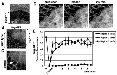
Barrier junctions prevent pathogen invasion and restrict paracellular leakage across epithelial sheets. To understand how one barrier junction, the septate junction (SJ), is regulated in vivo, we used fluorescence recovery after photobleaching (FRAP) to examine SJ protein dynamics in Drosophila. Most SJ-associated proteins, including Coracle, Neurexin IV and Nervana 2, displayed similar, extremely immobile kinetics. Loss of any of these components resulted in dramatically increased mobility of all others, suggesting that they form a single, highly interdependent core complex. Immobilization of SJ core components coincided with formation of the morphological SJ but occurred after their known role in maintaining epithelial polarity, suggesting that these functions are independent. In striking contrast to the core components, the tumor suppressor protein Discs large was much more mobile and its loss did not affect mobility of core SJ proteins, suggesting that it is not a member of this complex, even though it colocalizes with the SJ. Similarly, disruption of endocytosis affected localization of SJ core components, but did not affect their mobility. These results indicate that formation of a stable SJ core complex is separable from its proper subcellular localization, and provide new insights into the complex processes that regulate epithelial polarity and assembly of the SJ.


Figures

Fig. 1.
Fig. 1.
SJ core proteins display extremely slow FRAP rates. GFP-tagged proteins expressed in the lateral epidermis of stage 14 to 16 embryos were photobleached in the regions indicated (white ellipses). (A) Time-series images of photobleached regions for proteins indicated. Note that although Dlg–GFP recovers substantially within 5 minutes, little recovery of SJ core proteins was observed even after much longer time periods. Scale bars: 2 μm. (B) Average relative fluorescent recoveries of GFP-tagged SJ proteins. Dlg and E-cadherin, which display faster recoveries, are also plotted. E-cad–GFP was directly driven by ubiquitin promoter. Error bars indicate s.e.m. (C) Kymographs showing the recovery over time of a single bleached region of the membrane (taken from supplementary material Movies 1–5, 8–9). White arrowheads indicate time points of photobleaching. Black bars show initial width of the bleached regions. Note the overall lack of recovery for ATPα, Nrv2, Nrx-IV, Nrg, and Cora, as well as the minimal spreading of fluorescence from adjacent, unbleached regions of the membrane. By contrast, Dlg shows rapid recovery and spreading from lateral regions.
Fig. 2.
Fig. 2.
FLIP analysis of SJ core protein lateral mobility in the membrane. ATPα–GFP (A) and Nrx-IV-GFP (B) in the wild-type embryonic lateral epidermis, and Nrx-IV–GFP in nrv2 mutants (C) were continuously photobleached. Kymographs (right panels) were generated from the FLIP movies (left panels and supplementary material Movies 6, 7, 12). White lines indicate the bleached areas. The continuously bleached region spreads laterally for Nrx-IV in nrv2 mutant embryos, but not for ATPα or Nrx-IV in wild-type embryos. Scale bars: 1 μm.
Fig. 3.
Fig. 3.
Mobility of SJ proteins is not altered as cells divide. (A) GFP-tagged ATPα, Nrv2, Nrx-IV and Dlg in the columnar cells of wing discs were photobleached. White ellipses indicate bleached regions. (B) FRAP of SJ proteins in the columnar cells is plotted. These plots are similar to those for the embryonic epidermis. Error bars indicate s.e.m. (C) Nrx-IV–GFP was photobleached in a pair of mitotic columnar cells of the wing disc blade region. Fluorescent recovery in the cleavage furrow and polar region was observed (right panels), and relative fluorescence of this movie was plotted (left graph and supplementary material Movie 10). Scale bars: 1 μm (A), 2 μm (C).
Fig. 4.
Fig. 4.
Immobilization and localization of SJ core proteins occur coordinately during embryonic stage 13. (A) ATPα-GFP was photobleached (white ellipses) and then fluorescence recovery was monitored in stage 12 and 13 embryos. (B) Average of ATPα–GFP fluorescence recovery at each stage is plotted. Note that the onset of ATPα–GFP immobilization occurred in early stage 13. Error bars indicate s.e.m. (C) ATPα–GFP and Cora subcellular localization in stage 12–14 embryos. Vertical views of the lateral epidermis are shown. Arrowheads indicate specific accumulation of the SJ proteins. Dotted lines show the basal side. Note that immobilization and specific localization of ATPα–GFP coincided at stage 13. (D) Tangential views of ATPα-GFP in the embryonic lateral epidermis from stage 12 to 14. Starting at early stage 13, regions of locally more intense SJ protein localization are observed along the membrane (arrows). Scale bars: 1 μm (A), 5 μm (C), 2 μm (D).
Fig. 5.
Fig. 5.
Epidermal cells in stage 13 embryos individually acquire competency to form immobile SJs. (AC) Multiple contacts of two nearby epidermal cells expressing ATPα–GFP were bleached and recovery rates for each contact separately determined. All contacts in the ‘competent’ cell display less mobility than did those in the ‘incompetent’ cell. Numbered ellipses in A correspond to the positions plotted in C, which displays fluorescent recovery kinetics. Green and magenta lines display incompetent and competent cells, respectively. (DF) A competent epidermal cell can have both mobile and immobile contacts with adjacent cells. (D) Three distinct contacts in epidermal cells expressing ATPα–GFP were photobleached (ellipses marked 1–3). (E) Plots of the fluorescence recovery in regions shown in D. The magenta line indicates the area in which ATPα-GFP was stable, and green lines are the areas in which fluorescence recovered more quickly. (F) An interpretation based on the data in D,E, indicating competent and incompetent cells. Scale bars: 2 μm.
Fig. 6.
Fig. 6.
SJs fail to assemble on the border between cora-knockdown and wild-type cells. (A) RNAi-mediated knockdown of Cora expression. A portion of a wing imaginal disc expressing double-stranded cora RNA under the control of dpp-Gal4, stained with anti-Cora antibody. Note that Cora is still expressed in the thin peripodial cell layer that overlaps the cora-knockdown region. (B,C) Nrg–GFP in cora-depleted (top right) and wild-type cells (bottom left). Cytoplasmic fluorescence of GFPnls marks the cells expressing double-stranded cora RNA, whereas Nrg–GFP is strongly membrane associated. Boxed region is magnified in C. At the border between the intact and cora-knockdown cells, membrane-associated Nrg–GFP fluorescence is strongly decreased compared with levels in wild-type cells (arrowhead). (D) FRAP of Nrg–GFP in wild-type (region 3) and cora-depleted (region 2) cells, and on the border (region 1). (E) Average of Nrg–GFP fluorescence recovery in each region is plotted. To avoid measuring cytoplasmic GFPnls fluorescence, only fluorescence at the membrane was imaged. Error bars indicate s.e.m. Scale bars: 20 μm (A), 5 μm (B), 2 μm (D).
Fig. 7.
Fig. 7.
Loss of SJ components dramatically affects the mobility of SJ core proteins but not Dlg. FRAP of four GFP-tagged SJ proteins (ATPα, Nrv2, Nrx-IV and Nrg) and Dlg in zygotic SJ and endocytic mutant backgrounds in stage 14 or later embryos. Average of the fluorescent recovery of ATPα-GFP (A), Nrv2-GFP (B), Nrx-IV–GFP (C,E), Nrg–GFP (D) and Dlg–GFP (F) is plotted. Δvari (A) denotes Df(2L)Exel7079, ΔGli (C) denotes Df(2L)BSC255, Δcrim (D) denotes Df(3L)ED4470, and ΔAtpα and Atpα (D) denote the Df(3R)ED10811 homozygote and Df(3R)ED10811/Atpα01453a transheterozygote, respectively. Error bars indicate s.e.m.
Fig. 8.
Fig. 8.
Immobilization of SJ core proteins occurs independently of Dlg. (A,B) SJ proteins are mislocalized in the dlg maternal/zygotic (MZ) mutant embryos. Cora (A), PATJ (A) and ATPα–GFP (B) are all mislocalized in dlg MZ embryos. (C) Example of mobile and stable Nrv2–GFP at cell contacts in a stage 17 dlg MZ mutant. White arrowheads indicate ‘mobile’ contacts and the red arrow indicates a ‘stable’ contact. Areas in which fluorescence recovered to more than 60% within 7 minutes are regarded as ‘mobile’. (D) Recovery kinetics of ATPα–GFP, Nrv2–GFP and Nrx-IV–GFP in the dlg MZ mutant background. Error bars indicate s.e.m. Scale bars: 5 μm (A,B), 2 μm (C).
Fig. 9.
Fig. 9.
Schematic model of SJ maturation during embryogenesis. SJ proteins are initially highly mobile on the lateral membrane (stage 12), but in stage 13, Sinu, Mega, Cora, Vari, Nrx-IV, Nrg, Nrv2 and ATPα begin to form a stable SJ core complex just basal to the AJ. Four Ly6-like proteins (Bou, Crok, Cold and Crim) are necessary for this process. In addition, proper localization of the core complex requires Dlg, Gli and endocytosis. See Discussion for further details.'

Similar articles

Cited by

References

    1. Baumgartner S., Littleton J. T., Broadie K., Bhat M. A., Harbecke R., Lengyel J. A., Chiquet-Ehrismann R., Prokop A., Bellen H. J. (1996). A Drosophila neurexin is required for septate junction and blood-nerve barrier formation and function. Cell 87, 1059-1068 - PubMed
    1. Behr M., Riedel D., Schuh R. (2003). The claudin-like megatrachea is essential in septate junctions for the epithelial barrier function in Drosophila. Dev. Cell 5, 611-620 - PubMed
    1. Bhat M. A., Rios J. C., Lu Y., Garcia-Fresco G. P., Ching W., St Martin M., Li J., Einheber S., Chesler M., Rosenbluth J., et al. (2001). Axon-glia interactions and the domain organization of myelinated axons requires neurexin IV/Caspr/Paranodin. Neuron 30, 369-383 - PubMed
    1. Bilder D., Perrimon N. (2000). Localization of apical epithelial determinants by the basolateral PDZ protein Scribble. Nature 403, 676-680 - PubMed
    1. Bilder D., Li M., Perrimon N. (2000). Cooperative regulation of cell polarity and growth by Drosophila tumor suppressors. Science 289, 113-116 - PubMed

Publication types

MeSH terms