. 2011 Sep;9(9):1232-41.
doi: 10.1158/1541-7786.MCR-11-0098. Epub 2011 Aug 1.

Suppression of autophagy by FIP200 deletion impairs DNA damage repair and increases cell death upon treatments with anticancer agents

Affiliations

Suppression of autophagy by FIP200 deletion impairs DNA damage repair and increases cell death upon treatments with anticancer agents

Heekyong Bae et al. Mol Cancer Res. 2011 Sep.

Abstract

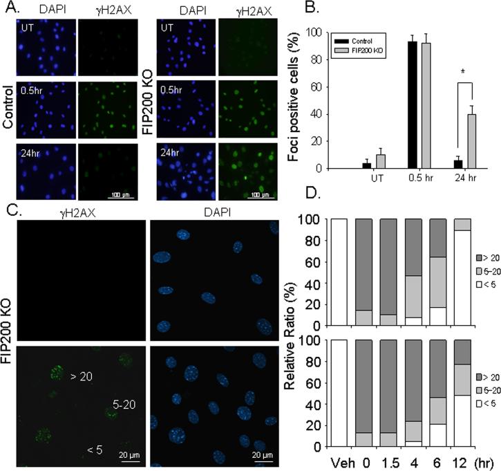
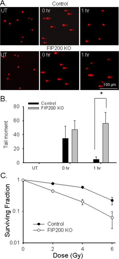
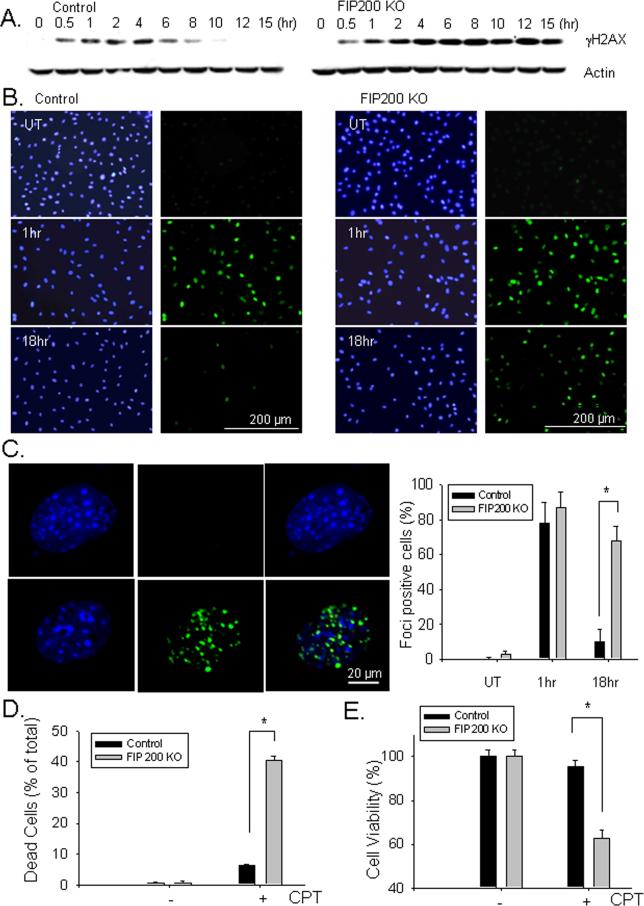
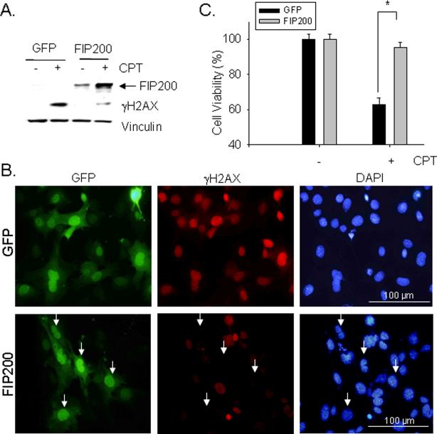
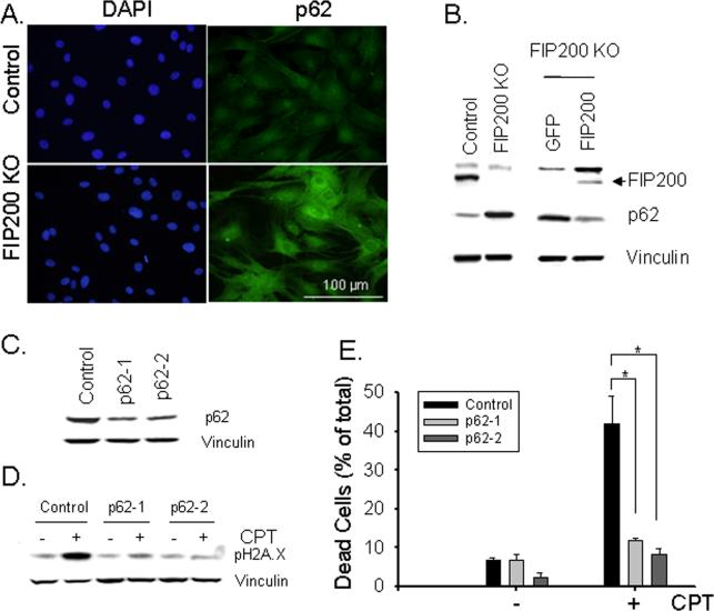
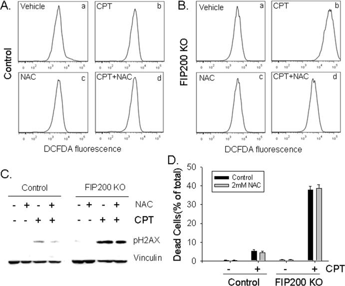
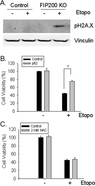
Autophagy is a lysosomal bulk degradation process for intracellular protein and organelles. FIP200 (200 kDa FAK-family interacting protein) is an essential component of mammalian autophagy that is implicated in breast cancer in recent studies. Here we show that inactivation of FIP200 resulted in deficient repair of DNA damage induced by ionizing radiation and anticancer agents in mouse embryonic fibroblasts (MEF). The persistent DNA damage correlated to increased apoptosis and reduced survival of FIP200 knockout (KO) MEFs after treatments with camptothecin (CPT), a topoisomerase I inhibitor and chemotherapeutic agent. Reexpression of FIP200 in FIP200 KO MEFs restored both efficient DNA damage repair and cell survival. Furthermore, knockdown of the increased p62 expression in FIP200 KO MEFs rescued the impaired DNA damage repair and CPT-induced cell death. In contrast, treatment of cells with N-acetyl cysteine did not affect these defects in FIP200 KO MEFs. Finally, FIP200 KO MEFs also showed deficient DNA damage repair and increased cell death compared with control MEFs, when treated with etoposide, a topoisomerase II inhibitor and another anticancer agent. Together, these results identify a new function for FIP200 in the regulation of DNA damage response and cell survival through its activity in autophagy and suggest the possibility of FIP200 or other autophagy proteins as a potential target for treatment to enhance the efficiency of cancer therapy using DNA damage-inducing agents.


Figures

Fig. 1
Fig. 1. Analysis of ionizing radiation-induced DNA damage and repair in FIP200 KO MEFs
(A-B) Control and FIP200 KO MEFs were exposed to 0 (UT) or 10 Gy of X-ray followed by 0.5 or 24 hr incubation at 37°C, as indicated. They were then fixed and immunostained with anti-γH2AX antibodies and DAPI (A). The percentage of cells with nuclear foci marked by γH2AX was determined under higher magnification. Mean ± SEM from three experiments are shown in panel B. *P<0.05. (C-D) Control and FIP200 KO MEFs were exposed to 0 (UT) or 1.5 Gy of X-ray followed by incubation at 37°C for various times, as indicated. The cells were then fixed, and immunostained with anti-γH2AX antibodies and DAPI, and viewed under higher magnification to visualize individual foci. Panel C shows representative images of cells with < 5, 5-20, or >20 foci. Panel D shows percentages of cells with <5, 5–20, or >20 foci per cell.
Fig. 2
Fig. 2. Analysis of ionizing radiation-induced DNA double strand break and cell survival in FIP200 KO MEFs
(A-B) Control and FIP200 KO MEFs were exposed to 0 (UT) or 10 Gy of X-ray followed by 0 or 1 hr incubation at 37°C, as indicated. They were then analyzed for DNA breaks by comet assay under neutral conditions. Representative images of cells with comet tails are shown in panel A. Tail moment was calculated by computerized image analysis with Comet Score™ software. Mean ± SEM from three experiments are shown in panel B. *P<0.05. (C) Control and FIP200 KO MEFs were exposed to various dose of IR, as indicated. Cells were then subjected to clonogenic survival assay as described in the Materials and Methods. The ratio between IR-treated and non-irradiated cells is shown. Results were from three independent experiments.
Fig. 3
Fig. 3. Analysis of DNA damage repair and cell survival in FIP200 KO MEFs after CPT treatment
(A). Control and FIP200 KO MEFs were treated with 5 μM CPT for various times, as indicated. The cells were then lyzed and analyzed by Western blotting using anti-γH2AX or anti-β-actin antibodies. (B and C) Control and FIP200 KO MEFs were treated with mock (UT) or 5 μM CPT for 1 or 18 hr as indicated. They were then fixed and immunostained with anti-γH2AX antibodies and DAPI (B). In Panel C, the cells were viewed under higher magnification to visualize individual foci. The percentage of cells with > 5 foci per cell was determined. Representative images are shown on the left and Mean ± SEM from three experiments are shown on the right. *P<0.05. (D and E) The percentage of dead cells at 48 hr after treatment with 5 μM CPT are determined by PI staining (D) and the cell viability was measured by MTT assay (E). Mean ± SEM from three experiments are shown. *P<0.05.
Fig. 4
Fig. 4. FIP200 re-expression rescues impaired DNA damage repair and cell survival in FIP200 KO MEFs
FIP200 KO MEFs were infected by recombinant adenoviruses encoding GFP or GFP-FIP200 fusion protein (FIP200) for two days, as indicated. (A) The cells were then treated with or without 5 μM CPT for 18 hr. The lysates were prepared and analyzed by Western blotting using various antibodies as indicated. (B) The infected cells were treated with 5 μM CPT for 18 hr and then examined by immunofluorescence using various antibodies as indicated. (C) The infected cells were incubated with 5 μM CPT for 48 hr and then cell viability was measured by MTT assay, as described in Fig. 3E. *P<0.05.
Fig. 5
Fig. 5. Increased expression of p62 and its role in the defective DNA damage repair and cell survival in FIP200 KO MEFs
(A) Control and FIP200 KO MEFs were fixed and immunostained with anti-p62 antibodies and DAPI. (B) Lysates were prepared from Control and FIP200 KO MEFs (left) or FIP200 KO MEFs that had been infected with recombinant adenoviruses encoding GFP or GFP-FIP200 fusion protein (FIP200) for two days (right). They were then analyzed by Western blotting using various antibodies as indicated. (C-E) FIP200 KO MEFs were infected by recombinant lentiviruses encoding shRNA targeting two different part of p62, or a scramble sequence as a control, as indicated. The cells were then incubated with 5 μM CPT for 18 hr (D, + lanes), 48 hr (E, + bars), or left untreated (C, - lanes and bars in D and E). Lysates were prepared and analyzed by Western blotting using various antibodies as indicated (C and D). The percentage of dead cells are determined by PI staining (E), as described in Fig. 3D. *P<0.05.
Fig. 6
Fig. 6. ROS scavenging does not affect the impaired DNA damage repair and reduced survival of FIP200 KO MEFs after CPT treatment
(A and B) Control and FIP200 KO MEFs were treated with 5 μM CPT, 2 mM NAC, or both, as indicated. They were then stained by DCFDA and analyzed by FACS, as described in the Materials and Methods. Representative FACS profiles of DCFDA staining are shown. (C and D) Control and FIP200 KO MEFs were pretreated with 2 mM NAC for 45 min, and then incubated with 5 μM CPT for 18 hr (C) or 48 hr (D), as indicated. Lysates were prepared and analyzed by Western blotting using various antibodies as indicated (C). The percentage of dead cells are determined by PI staining (D), as described in Fig. 3D. *P<0.05.
Fig. 7
Fig. 7. Analysis of DNA damage repair and cell survival in response to etoposide
(A) Control and FIP200 KO MEFs were stimulated with 25 μM etoposide for 24 hr. Cell lysates were prepared and analyzed by Western blotting using various antibodies as indicated. (B) FIP200 KO MEFs were infected by recombinant lentiviruses encoding shRNA targeting p62, or a scramble sequence as a control along with a puromycin-resistant marker, as indicated. The infected cells were selected in 5 μg/ml puromycin for two days, and then treated with or without 25 μM etoposide for two days. Cell viability was measured by MTT assay, as described in Fig. 3E. *P<0.05. (C) FIP200 KO MEFs were pretreated with 2 mM NAC for 45 min, and then incubated with 25 μM etoposide for two days, as indicated. Cell viability was measured by MTT assay, as described in Fig. 3E.

Similar articles

Cited by

References

    1. Simonsen A, Tooze SA. Coordination of membrane events during autophagy by multiple class III PI3-kinase complexes. 2009. pp. 773–82. - PMC - PubMed
    1. He C, Klionsky DJ. Regulation mechanisms and signaling pathways of autophagy. Annual review of genetics. 2009;43:67–93. - PMC - PubMed
    1. Rabinowitz JD, White E. Autophagy and metabolism. Science. 330(6009):1344–8. - PMC - PubMed
    1. Kroemer G, Marino G, Levine B. Autophagy and the integrated stress response. Mol Cell. 40(2):280–93. - PMC - PubMed
    1. Levine B, Kroemer G. Autophagy in the pathogenesis of disease. Cell. 2008;132(1):27–42. - PMC - PubMed

Publication types

MeSH terms