. 2011 Aug 16;108(33):13641-6.
doi: 10.1073/pnas.1108440108. Epub 2011 Aug 1.

Genetic framework for GATA factor function in vascular biology

Affiliations

Genetic framework for GATA factor function in vascular biology

Amelia K Linnemann et al. Proc Natl Acad Sci U S A. .

Abstract

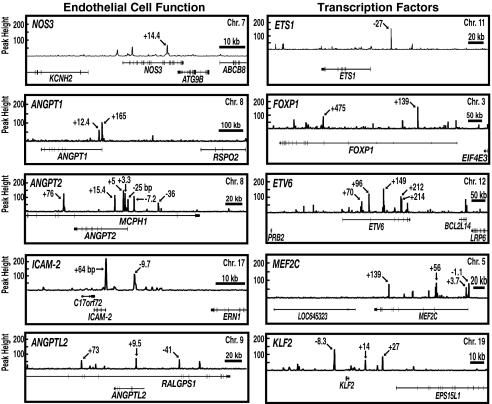
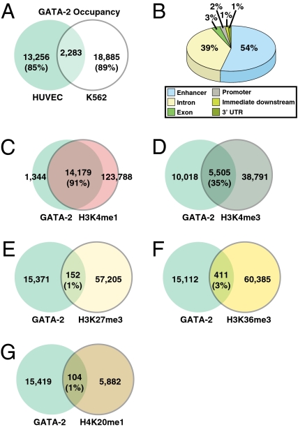
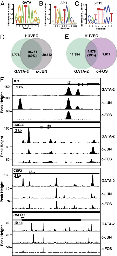
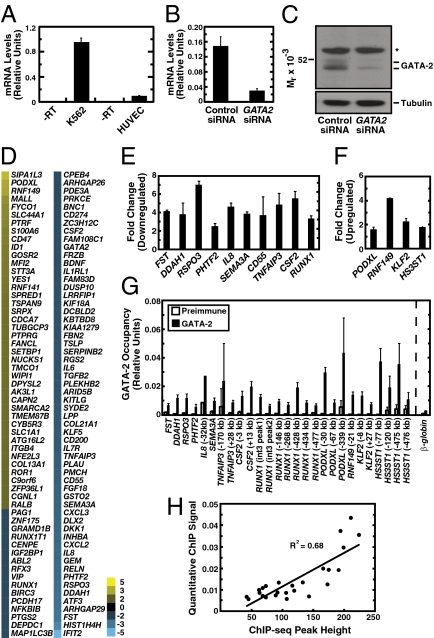
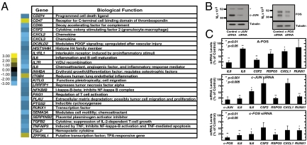
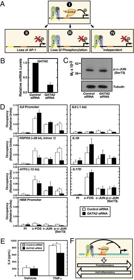
Vascular endothelial dysfunction underlies the genesis and progression of numerous diseases. Although the GATA transcription factor GATA-2 is expressed in endothelial cells and is implicated in coronary heart disease, it has been studied predominantly as a master regulator of hematopoiesis. Because many questions regarding GATA-2 function in the vascular biology realm remain unanswered, we used ChIP sequencing and loss-of-function strategies to define the GATA-2-instigated genetic network in human endothelial cells. In contrast to erythroid cells, GATA-2 occupied a unique target gene ensemble consisting of genes encoding key determinants of endothelial cell identity and inflammation. GATA-2-occupied sites characteristically contained motifs that bind activator protein-1 (AP-1), a pivotal regulator of inflammatory genes. GATA-2 frequently occupied the same chromatin sites as c-JUN and c-FOS, heterodimeric components of AP-1. Although all three components were required for maximal AP-1 target gene expression, GATA-2 was not required for AP-1 chromatin occupancy. GATA-2 conferred maximal phosphorylation of chromatin-bound c-JUN at Ser-73, which stimulates AP-1-dependent transactivation, in a chromosomal context-dependent manner. This work establishes a link between a GATA factor and inflammatory genes, mechanistic insights underlying GATA-2-AP-1 cooperativity and a rigorous genetic framework for understanding GATA-2 function in normal and pathophysiological vascular states.


Conflict of interest statement

The authors declare no conflict of interest.

Figures

Fig. 1.
Fig. 1.
Representative GATA-2 target genes in HUVEC. Signal maps of GATA-2 occupancy sites in HUVEC identified by ChIP-seq. Genes important for endothelial cell function and transcription factors are depicted. Arrows indicate ChIP-seq peak locations relative to the transcription start site of the respective GATA-2 target gene (kb).
Fig. 2.
Fig. 2.
Computational mining of ChIP-seq data. (A) Comparison of GATA-2–occupied peaks in HUVEC and K562 cells. (B) Locations of GATA-2–occupied peaks relative to nearest-neighbor genes determined with the Cis Element Annotation System (http://liulab.dfci.harvard.edu/CEAS/; http://ceas.cbi.pku.edu.cn/). (CG) Comparison of GATA-2–occupied peaks and peaks of H3K4me1 (C), H3K4me3 (D), H3K27me3 (E), H3K36me3 (F), and H4K20me1 (G).
Fig. 3.
Fig. 3.
Linking GATA-2 and AP-1 function. (AC) Logos of overrepresented motifs from GATA-2–occupied ChIP-seq peaks: (A) GATA motif from constrained motif analysis and (B) AP-1 and (C) c-ETS motifs from de novo motif finding. (D and E) Comparison of GATA-2–occupied peaks and peaks of (D) c-JUN and (E) c-FOS occupancy in HUVEC. (F) Representative profiles demonstrating overlap among GATA-2, c-JUN, and c-FOS occupancy peaks.
Fig. 4.
Fig. 4.
Direct GATA-2 target gene ensemble. (A) Comparison of GATA2 levels in HUVEC and K562 cells. (B) Quantitative RT-PCR analysis of GATA2 transcript levels after treatment with nontargeting control siRNA or GATA2 siRNA. (C) Western blotting of GATA-2 in HUVEC transfected with control or GATA2 siRNA. Whole-cell samples were analyzed. *, nonspecific band. (D) Heat map depicting the mean fold change of expression at GATA-2–occupied loci resulting from GATA2 knockdown (n = 2). (E and F) Quantitative RT-PCR validation of array results in GATA-2–activated (down-regulated with knockdown) (E) and repressed (up-regulated) genes (F). (G) Quantitative ChIP validation of GATA-2 occupancy at loci dysregulated by the knockdown. Data shown are mean ± SE; n = 3. (H) Correlation between height of ChIP-seq peak and quantitative ChIP signal.
Fig. 5.
Fig. 5.
GATA-2/AP-1–dependent regulation of inflammatory genes. (A) Genes involved in inflammation and their expression changes with GATA-2 knockdown. Blue, down-regulated with GATA-2 knockdown; Yellow, up-regulated with GATA-2 knockdown. (B) Western blotting of endogenous c-JUN and c-FOS in HUVEC transfected with c-JUN or c-FOS siRNA, respectively. Transfected cells were boiled in SDS-sample buffer, and whole-cell samples were analyzed by Western blotting. *, nonspecific band. (C) Real-time RT-PCR analysis of gene expression in HUVEC transfected with a dominant-negative AP-1 antagonist (A-FOS), c-JUN siRNA, or c-FOS siRNA. The expression of empty vector-transfected cells (A-FOS) or nontargeting control siRNA-transfected cells (c-JUN and c-FOS) was designated as 100%, and expression for each gene is represented as a percentage of control expression. Data shown are mean ± SE; n ≥ 3.
Fig. 6.
Fig. 6.
GATA-2 requirement for assembly of functional AP-1 complexes on chromatin. (A) Models as described in Results and Discussion. (B) Real-time RT-PCR quantitation of GATA2 mRNA. (C) Western blotting of c-JUN phosphorylated at serine 73 [p-c-JUN (Ser73)] in HUVEC transfected with control or GATA2 siRNA. Whole-cell samples were analyzed by Western blotting. (D) Quantitative ChIP of preimmune control (PI), c-FOS, c-JUN, and p-c-JUN (Ser-73) occupancy at the IL-8 promoter, RSPO3, and ATF3, AP-1 targets that are GATA-2–independent (IL-1B, IL-17D), and negative controls (HBB and a site 1 kb upstream of the IL-8 promoter). Data shown are mean ± SE; n ≥ 3. (E) ELISA quantitation of IL-8 levels in medium from control siRNA- or GATA-2 siRNA-transfected cells treated with 10 ng/mL TNF-α or vehicle for 6 h. Data shown are mean ± SE; n = 6. (F) GATA-2–AP-1 coregulation of inflammatory genes in endothelium.

References

    1. De Val S, Black BL. Transcriptional control of endothelial cell development. Dev Cell. 2009;16:180–195. - PMC - PubMed
    1. Tsai F-Y, Orkin SH. Transcription factor GATA-2 is required for proliferation/survival of early hematopoietic cells and mast cell formation, but not for erythroid and myeloid terminal differentiation. Blood. 1997;89:3636–3643. - PubMed
    1. Tsai FY, et al. An early haematopoietic defect in mice lacking the transcription factor GATA-2. Nature. 1994;371:221–226. - PubMed
    1. Bresnick EH, Lee HY, Fujiwara T, Johnson KD, Keles S. GATA switches as developmental drivers. J Biol Chem. 2010;285:31087–31093. - PMC - PubMed
    1. Dorfman DM, Wilson DB, Bruns GA, Orkin SH. Human transcription factor GATA-2. Evidence for regulation of preproendothelin-1 gene expression in endothelial cells. J Biol Chem. 1992;267:1279–1285. - PubMed

Publication types

Associated data