. 2011 Aug 16;108(33):13552-7.
doi: 10.1073/pnas.1016778108. Epub 2011 Aug 1.

Role for the actomyosin complex in regulated exocytosis revealed by intravital microscopy

Affiliations

Role for the actomyosin complex in regulated exocytosis revealed by intravital microscopy

Andrius Masedunskas et al. Proc Natl Acad Sci U S A. .

Abstract

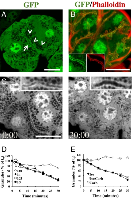
The regulation and the dynamics of membrane trafficking events have been studied primarily in in vitro models that often do not fully reflect the functional complexity found in a living multicellular organism. Here we used intravital microscopy in the salivary glands of live rodents to investigate regulated exocytosis, a fundamental process in all of the secretory organs. We found that β-adrenergic stimulation elicits exocytosis of large secretory granules, which gradually collapse with the apical plasma membrane without any evidence of compound exocytosis, as was previously described. Furthermore, we show that the driving force required to complete the collapse of the granules is provided by the recruitment of F-actin and nonmuscle myosin II on the granule membranes that is triggered upon fusion with the plasma membrane. Our results provide information on the machinery controlling regulated secretion and show that intravital microscopy provides unique opportunities to address fundamental questions in cell biology under physiological conditions.


Conflict of interest statement

The authors declare no conflict of interest.

Figures

Fig. 1.
Fig. 1.
A transgenic mouse model for dynamic imaging of the SCGs the APM in vivo. The submandibular SGs of anesthetized GFP mice were imaged in situ by IVM (A and C) or excised and labeled with TRITC–phalloidin to reveal the actin cytoskeleton (B). (A) Cytoplasmic GFP (green) is excluded from the SCGs (arrowheads) and enriched at the APM (arrow). (B) The APM is enriched in GFP (green) and actin as revealed by counterstaining with TRITC–phalloidin (red). (Insets) High magnification of a canaliculus. (C) The anesthetized GFP mice received a s.c. injection of 0.5 mg/kg Iso, and the SGs were imaged by time-lapse confocal microscopy. Acinus imaged at the moment of Iso injection (Left) or after 30 min (Right) (Movies S2 and S3). High magnification of the canaliculi (Insets). (Scale bars, 10 μm.) (D and E) Quantitation of the degranulation of the SCGs. Data shown are measurements from a single acinus. The experiments were performed three times with similar results. (D) Exocytosis in response to various doses of Iso: 0.01 mg/kg (○), 0.1 mg/kg (●), 0.25 mg/kg (□), and 0.5 mg/kg (■). (E) Exocytosis in response to muscarinic or adrenergic stimulation: 0.01 mg/kg Carb (○), 0.1 mg/kg Iso (●) or both (□).
Fig. 2.
Fig. 2.
SCGs completely collapse after fusion with the APM. (A) SCGs close to the APM are enriched in GFP. Anesthetized GFP mice were injected s.c. with 0.1 mg/kg Iso, and after 10 min, excised and labeled with TRITC–phalloidin. Granules close to the APM are coated with actin (red) and also enriched in GFP (green). (Scale bar, 10 μm.) (B) Time series of a single granule (white arrows) fusing with the APM (red line) (red arrowheads in Movie S6). (Scale bar, 3 μm.) (C) Quantitation of both the GFP fluorescence intensity around a SCG (black circles) and its diameter (red circles) during Iso-stimulated exocytosis. Data shown are from a single event. Five events per animal were measured in two independent experiments. (DF) Texas Red–dextran (10 kDa) was infused by gravity into the salivary ducts of a live anesthetized rat. The SGs were exposed and imaged by confocal microscopy. (D) Canaliculi are highlighted in the acini (arrows). (Inset) High magnification of a canaliculus. (Scale bar, 10 μm.) (E and F) Iso (0.1 mg/kg) was injected s.c. (E) Time-lapse sequence. When the fusion pore opens, dextran enters two SCGs (white and red arrows), enabling their visualization. (Scale bar, 3 μm.) (Movie S7). (F) The fluorescence intensity of the dextran inside the granule shown in E (white arrows) was measured (black circles) and correlated with its diameter (red circles) as described in Materials and Methods. Data shown are from a single event. Four to five events per animal were measured in two independent experiments. (GJ) Anesthetized m-Tomato mice were left untreated (G and I) or injected with Iso (H, J, and K). The SMGs were imaged in situ (G, H, and J) or labeled with TRITC–phalloidin (I). Acinar canaliculi in cross-section [G, H (Inset), and I]. (J) Time series of the collapse of two SCGs revealed by the diffusion of the m-Tomato from the APM (white and red arrows). Note the expansion of the canaliculi (white and red arrowheads). (Scale bar, 3 μm.) (Movie S9). (K) The fluorescence intensity of the m-Tomato at the surface of a granule (black circles) and its diameter (red circles) were measured as described in Materials and Methods. Data shown are measurements of a single event. A total of 6–10 events per animal were recorded in three independent experiments.
Fig. 3.
Fig. 3.
Role of the actin cytoskeleton in facilitating the collapse of the SCGs. (AD) The SGs of the m-Tomato mice were exposed to 10 μM CD, and after 20 min the animals received a s.c.injection of 0.1 mg/kg Iso. (A) The SGs were exposed and imaged in situ. Large vacuoles were observed in the cytoplasm of the acinar cells (arrows). (Scale bar, 10 μm.) (BD) Time-lapse sequence of SCGs after fusion with the APM, 2 min (B) or 10 min (D) after the injection of Iso. (B) The SCGs fuse with the APM, do not collapse, and form large vacuoles. (Scale bar, 3 μm.) (Movie S12). (C) Quantitation of the diameter of the vacuoles that fail to collapse after fusion with the APM in the presence of CD (red circles) and of the granules that fuse under control conditions (black circles). Data shown are from four granules in the same acinus. A total of 10–15 granules were analyzed in two independent experiments. (D) A SCG fuses with a large vacuole and rapidly collapses (arrows) (Movie S14).
Fig. 4.
Fig. 4.
Role of myosin IIa and IIb in the collapse of the secretory granules. (AD) Friend Virus B-Type (FVB) mice were left untreated (A and C) or injected with 0.1 mg/kg Iso (B and D). SGs were excised and labeled for endogenous nonmuscle myosin IIa (A and B) and IIb (C and D) and actin (B and C) as described in Materials and Methods. Upon stimulation with Iso, both myosin types are localized in SCGs at the APM (arrowheads). (Inset in B) An SCG at the APM. (Scale bars, 10 μm.) (EH) The SGs of m-Tomato mice were exposed and incubated with 50 μM of either (−)Bleb (E and G) or (+)Bleb (F and H). After 20 min, the SGs were imaged (E and F, Left panels). In the presence of (−)Bleb, the acinar canaliculi expanded (arrows). The mice were then injected s.c. with 0.1 mg/kg Iso and imaged in time-lapse mode (E and F, Right panels, and G and H). (E and F) Snapshots taken 10 min after the Iso injection. Large granules appeared at the APM (arrowheads). (Scale bar in F, 10 μm.) (G and H) Time-lapse sequence of SCGs fusing with APM (Movies S15 and S16). Expanded canaliculus (asterisk). (Scale bars, 3 μm.) (I) Quantitation of the diameter of the SCGs after fusion with the APM in the presence of (−)Bleb (red circles) or (+)Bleb (black circles). Data shown are from four granules in the same acinus. A total of 15–20 granules were analyzed in two independent experiments.

Similar articles

Cited by

References

    1. Burgoyne RD, Morgan A. Secretory granule exocytosis. Physiol Rev. 2003;83:581–632. - PubMed
    1. De Matteis MA, Luini A. Exiting the Golgi complex. Nat Rev Mol Cell Biol. 2008;9:273–284. - PubMed
    1. Sudhof TC. The synaptic vesicle cycle. Annu Rev Neurosci. 2004;27:509–547. - PubMed
    1. Sokac AM, Bement WM. Kiss-and-coat and compartment mixing: Coupling exocytosis to signal generation and local actin assembly. Mol Biol Cell. 2006;17:1495–1502. - PMC - PubMed
    1. Castle AM, Huang AY, Castle JD. The minor regulated pathway, a rapid component of salivary secretion, may provide docking/fusion sites for granule exocytosis at the apical surface of acinar cells. J Cell Sci. 2002;115:2963–2973. - PubMed

Publication types

LinkOut - more resources