. 2011 Aug 16;108(33):13764-9.
doi: 10.1073/pnas.1111093108. Epub 2011 Aug 1.

Autism-linked neuroligin-3 R451C mutation differentially alters hippocampal and cortical synaptic function

Affiliations

Autism-linked neuroligin-3 R451C mutation differentially alters hippocampal and cortical synaptic function

Mark Etherton et al. Proc Natl Acad Sci U S A. .

Abstract

Multiple independent mutations in neuroligin genes were identified in patients with familial autism, including the R451C substitution in neuroligin-3 (NL3). Previous studies showed that NL3(R451C) knock-in mice exhibited modestly impaired social behaviors, enhanced water maze learning abilities, and increased synaptic inhibition in the somatosensory cortex, and they suggested that the behavioral changes in these mice may be caused by a general shift of synaptic transmission to inhibition. Here, we confirm that NL3(R451C) mutant mice behaviorally exhibit social interaction deficits and electrophysiologically display increased synaptic inhibition in the somatosensory cortex. Unexpectedly, however, we find that the NL3(R451C) mutation produced a strikingly different phenotype in the hippocampus. Specifically, in the hippocampal CA1 region, the NL3(R451C) mutation caused an ∼1.5-fold increase in AMPA receptor-mediated excitatory synaptic transmission, dramatically altered the kinetics of NMDA receptor-mediated synaptic responses, induced an approximately twofold up-regulation of NMDA receptors containing NR2B subunits, and enhanced long-term potentiation almost twofold. NL3 KO mice did not exhibit any of these changes. Quantitative light microscopy and EM revealed that the NL3(R451C) mutation increased dendritic branching and altered the structure of synapses in the stratum radiatum of the hippocampus. Thus, in NL3(R451C) mutant mice, a single point mutation in a synaptic cell adhesion molecule causes context-dependent changes in synaptic transmission; these changes are consistent with the broad impact of this mutation on murine and human behaviors, suggesting that NL3 controls excitatory and inhibitory synapse properties in a region- and circuit-specific manner.


Conflict of interest statement

The authors declare no conflict of interest.

Figures

Fig. 1.
Fig. 1.
NL3R451C knock-in mice exhibit social interaction impairments. (A) Three-chamber sociability test. Mice were first simultaneously exposed to an empty round wire container (pencil cup) as a novel object and a caged, unfamiliar mouse (Left). Afterward, a novel mouse was introduced into the empty wire container (Right). The sniffing duration for each mouse was measured as shown (paired t test; Left: WT = P < 0.01, R451C = P < 0.0001; Right: WT = P < 0.05, R451C = n.s.). (B) Caged adult social interaction test. Mice were sequentially exposed to a novel object (empty rectangular wired cage) and a caged stranger mouse, and the time course of interaction was measured in 30-s time bins. No differences in the genotype were detected during the novel object interaction trial (Upper) but were measured during the social trial (Lower; effect of genotype: F1, 306 = 4.47, P = 0.042). (C) Summary graphs of the total interaction time measured in B. (*P < 0.05 by unpaired t test; n.s. = nonsignificant). Data represent means ± SEMs.
Fig. 2.
Fig. 2.
NL3R451C knock-in mutation increases excitatory synaptic transmission in the CA1 region of the hippocampus. (A) Representative traces and summary graph of input–output measurements performed by extracellular field recordings in acute hippocampal slices from littermate WT and NL3R451C mutant mice (R451C). (B) Same as A, except that NL3 KO mice were analyzed. (C) Summary graph of the linear slope fits measured during input–output recordings for NL3 KO and NL3R451C mutant mice. Note that two independent lines of NL3R451C mutant mice were examined (the lines described by Tabuchi et al. in ref. , referred to as R451C, and Chadman et al. in ref. , referred to as R451C*) in addition to the NL3 KO mice. Data represent means ± SEMs. Statistical significance (P < 0.01) was evaluated with one-way ANOVA (A and B) or Student t test (C). Total number of slices and mice examined are shown in the bars of panel C. Paired-pulse measurements are in Fig. S1.
Fig. 3.
Fig. 3.
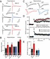
NL3R451C knock-in mutation increases NMDA receptor-mediated synaptic responses and LTP in the hippocampus. (A) Sample traces of measurements of the ratio of NMDA vs. AMPA receptor-mediated synaptic currents. Analyses compared littermate WT and NL3R451C mutant mice in the hippocampus and layer 2/3 of the somatosensory cortex and littermate WT and NL3 KO mice in the hippocampus as indicated. AMPA and NMDA receptor-mediated responses were monitored with postsynaptic holding potentials of −70 and +40 mV, respectively. (B) Summary graphs of the NMDA/AMPA receptor response ratios as indicated. Note that, as in Fig. 1, two independent lines of NL3R451C mutant mice were examined [in Tabuchi et al. (15), mice were referred to as R451C, and in Chadman et al. (23), mice referred to as R451C*]. Parallel measurements of spontaneous release are in Fig. S2. (C) Sample traces from extracellular field recordings performed in the CA1 region in acute slices of the hippocampus from littermate WT and NL3R451C mutant (Left) or WT and NL3 KO mice (Right). LTP was induced by three 1-s, 100-Hz stimulations. Traces are from before (1) or 60 min after LTP induction (2). (D) Summary graphs of the fEPSP slope as a function of LTP induction monitored in acute slices from WT and mutant mice as indicated. (E) Plot of the average percentage of synaptic transmission increase during LTP measured 55–60 min postinduction relative to baseline. Data represent means ± SEM; in B and E, total numbers of cells or slices/total number of mice are indicated in the bars, respectively. Statistical significance was evaluated with Student t test (*P < 0.05; **P < 0.01).
Fig. 4.
Fig. 4.
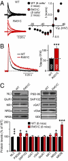
NL3R451C mutation alters kinetics and composition of synaptic NMDA receptors. (A) Normalized sample traces (Left) and summary graph (Right) for NMDA receptor current voltage measurements. (B) Normalized sample traces (Left) and summary graph (Right) for NMDA receptor decay kinetics measurements. (C) Representative immunoblots of the indicated proteins (Upper) and total protein levels in hippocampal lysates measured using quantitative immunoblotting with 125I-labeled antibodies and phosphoImager detection (Lower). Hippocampal lysates were from littermate WT and NL3R451C knock-in mice at 6 wk of age. Protein levels were normalized first to GDP-dissociation inhibitor (GDI) as an internal loading control and then, to WT levels. Data shown are means ± SEMs (*P < 0.05; ***P < 0.001 using Student t test; in B, total number of cells/total number mice analyzed are shown in the bar diagrams).
Fig. 5.
Fig. 5.
Changes in dendritic branching and synapse structure in NL3R451C mutant stratum radiatum of the CA1 region of the hippocampus. (A and B) Representative images (A) and summary graph of the number of dendritic branch points (B) of biocytin-filled pyramidal neurons in the CA1 region of the hippocampus. Neurons were filled with biocytin through a patch pipette in acute slices (str. lac. molecular, stratum lacunosum moleculare). (C) Representative images and summary graph of the spine density of dendrites in the stratum radiatum of Alexa-555–filled pyramidal neurons in the CA1 region of the hippocampus. (Scale bar: 1.985 μm.) (D) Representative electron micrographs from the stratum radiatum of the CA1 region of the hippocampus. (Scale bar: 1 μm.) (E and F) Summary graph of PSD size and number of docked vesicles in excitatory synapses of the stratum radiatum of the CA1 region of the hippocampus from littermate WT and NL3R451C mutant mice. (G–I) Quantitation of the presynaptic nerve terminal size (G), number of vesicles per terminal (H), and spine size (I) in excitatory synapses of the stratum radiatum of the CA1 region from littermate WT and NL3R451C mutant mice. Data represent means ± SEM. For B and C, numbers in bars indicate numbers of neurons/mice analyzed; for E–I, 303 electron micrographs from two pairs of littermates with 552 (WT) and 778 synapses (R451C) were measured. Statistical significance was evaluated by Student t test (*P < 0.05; **P < 0.01; ***P < 0.001).

References

    1. American Psychiatric Association . Diagnostic Criteria from DSM-IV-TR. Washington, DC: American Psychiatric Association; 2000.
    1. Folstein S, Rutter M. Infantile autism: A genetic study of 21 twin pairs. J Child Psychol Psychiatry. 1977;18:297–321. - PubMed
    1. Persico AM, Bourgeron T. Searching for ways out of the autism maze: Genetic, epigenetic and environmental clues. Trends Neurosci. 2006;29:349–358. - PubMed
    1. Jamain S, et al. Mutations of the X-linked genes encoding neuroligins NLGN3 and NLGN4 are associated with autism. Nat Genet. 2003;34:27–29. - PMC - PubMed
    1. Laumonnier F, et al. X-linked mental retardation and autism are associated with a mutation in the NLGN4 gene, a member of the neuroligin family. Am J Hum Genet. 2004;74:552–557. - PMC - PubMed

Publication types