. 2011 Aug 16;108(33):13480-5.
doi: 10.1073/pnas.1105959108. Epub 2011 Aug 1.

A bacterial process for selenium nanosphere assembly

Affiliations

A bacterial process for selenium nanosphere assembly

Charles M Debieux et al. Proc Natl Acad Sci U S A. .

Abstract

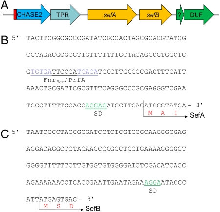
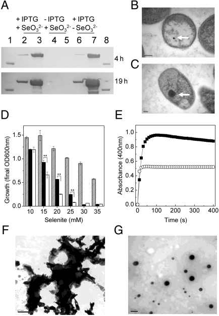
During selenate respiration by Thauera selenatis, the reduction of selenate results in the formation of intracellular selenium (Se) deposits that are ultimately secreted as Se nanospheres of approximately 150 nm in diameter. We report that the Se nanospheres are associated with a protein of approximately 95 kDa. Subsequent experiments to investigate the expression and secretion profile of this protein have demonstrated that it is up-regulated and secreted in response to increasing selenite concentrations. The protein was purified from Se nanospheres, and peptide fragments from a tryptic digest were used to identify the gene in the draft T. selenatis genome. A matched open reading frame was located, encoding a protein with a calculated mass of 94.5 kDa. N-terminal sequence analysis of the mature protein revealed no cleavable signal peptide, suggesting that the protein is exported directly from the cytoplasm. The protein has been called Se factor A (SefA), and homologues of known function have not been reported previously. The sefA gene was cloned and expressed in Escherichia coli, and the recombinant His-tagged SefA purified. In vivo experiments demonstrate that SefA forms larger (approximately 300 nm) Se nanospheres in E. coli when treated with selenite, and these are retained within the cell. In vitro assays demonstrate that the formation of Se nanospheres upon the reduction of selenite by glutathione are stabilized by the presence of SefA. The role of SefA in selenium nanosphere assembly has potential for exploitation in bionanomaterial fabrication.


Conflict of interest statement

The authors declare no conflict of interest.

Figures

Fig. 1.
Fig. 1.
Physiological analysis of Se-nanosphere production. (A) Growth curve of T. selenatis grown on acetate using selenate (10 mM) as the sole electron acceptor (Error bars are SEM; n = 10 cultures). Time points t1t6 indicate the samples used for EM analysis. (B) Transmission electron micrographs of time points from A. Micrographs t1 and t2 show midexponential phase, t3 and t4 show late exponential phase, and t5 and t6 show stationary phase. Scale bar, 200 nm. Selenium deposits are indicated by an arrow. Poly-β-hydroxybutarate granular deposits are indicated by an asterisk. (C and D) Transmission electron micrographs of purified Se nanospheres. (C) Scale bar, 500 nm. (D) Scale bar, 50 nm.
Fig. 2.
Fig. 2.
Protein analysis of Se nanospheres from T. selenatis. (A) SDS-PAGE gels stained for secreted proteins from T. selenatis grown under anaerobic conditions. Lane 1, Invitrogen See Blue Plus2 Prestained Standard; lane 2, protein from cells grown on selenate (10 mM); lane 3, protein from cells grown on nitrate (10 mM) plus selenite (10 mM). (B) SDS-PAGE gel stained for total protein secreted in the extracellular medium from cells grown aerobically under different growth conditions. Lane 1, Invitrogen See Blue Plus2 Prestained Standard; lane 2, control (cells grown in LB medium only); lane 3, LB medium containing 10 mM selenite prior to inoculation with T. selenatis; lane 4, cells grown in LB medium supplemented with 10 mM selenate; lane 5, cells grown in LB medium supplemented with 10 mM nitrate; and lane 6, cells grown in LB medium supplemented with 10 mM selenite. (C) Secreted proteins from T. selenatis grown under aerobic conditions on LB medium supplemented with selenite (10 mM) following incubation for 16, 24, and 40 h, respectively. (D) Analysis of protein secretion and regulation upon exposure to increasing concentrations of selenite. (i) Observed selenium precipitation in cultures, (ii) SDS-PAGE analysis of secreted proteins, (iii and iv) end-point RT-PCR of sefA and 16S transcripts, and (v) Northern blot of sefA transcript.
Fig. 3.
Fig. 3.
Analysis of the sef operon. (A) Schematic representation of the sef gene locus. Putative annotations are as follows: CHASE2 extracellular sensory domain and guanylate cyclase (with sec leader peptide in red); SefA, selenium nanosphere assembly protein; SefB, SAM-methyltransferase;?, putative peptide. The (B) nucleotide sequence of the promoter region of sefA. (C) Nucleotide sequence upstream from sefB. A putative Shine–Dalgarno (SD) sequence and putative FnrBac/PrfA binding motif (TGTGA-N6-TCACA) are located upstream of sefA. No obvious promoter binding sequences are identified between sefA and sefB. A putative SD sequence upstream of sefB is also located.
Fig. 4.
Fig. 4.
Expression of SefA in E. coli and in vitro formation of Se nanospheres. (A) Western blot analysis of the localization of SefA expression in E. coli following exposure to selenite. Samples (10 μg) were prepared from cells +/- IPTG and +/- selenite (10 mM). Lanes 2, 4, and 6 represent extracellular protein; lanes 3, 5, and 7 represent soluble cell extracts. Samples were analyzed at both 4 and 19 h after the addition of IPTG and selenite. Lanes 1 and 8 show an 80-kDa marker. (B) Transmission electron micrograph of an E. coli single cell harboring plasmid pET33b-sefA grown in the presence of selenite (10 mM), but not induced with IPTG. (C) Transmission electron micrograph of an E. coli single cell harboring plasmid pET33b-sefA grown in the presence of both selenite (10 mM) and IPTG. Three replicate cells are shown in Fig. S3. Selenium deposits are indicated by an arrow. (D) Growth yield at stationary phase of E. coli cells exposed to increasing selenite concentrations. E. coli harboring plasmid pET33b-sefA in the absence (hashed bars) or presence (solid bars) of IPTG, and E. coli cells harboring plasmid pET33b in the presence of IPTG (open bars). Error bars are SEM (n = 3). ** indicates t test, p < 0.01 compared to the nonrecombinant (pET33b) control. (E) The reaction of GSH (4 mM) with selenite (0.5 mM), either in the presence (○) or absence (▪) of purified rSefA (0.5 μg), monitored at 400 nm. (F and G) Transmission electron micrographs of the reaction product from E, in the absence (F) and in the presence (G) of SefA. Scale bars, 0.5 μm and 200 nm for F and G, respectively.
Fig. 5.
Fig. 5.
Schematic diagram showing the proposed pathway of selenium oxyanion reduction and Se-nanosphere assembly in T. selenatis. The reduction of selenate draws electrons from the membrane-bound QCR, generating a net gain of 2q+/2e- of proton electrochemical gradient, which could provide the driving force for the translocation of selenite across the cytoplasmic membrane. Once in the cytoplasm, selenite reduction occurs, and the resultant Se0 binds to SefA, forming a Se nanosphere prior to export from the cell. The process by which SefA–Se is exported remains unknown. The identification of a gene (sefB) encoding a putative SAM-dependent methyltransferase might also provide a mechanism for selenite detoxification via volatilization to methylated selenides (R–Se–R). OM, outer membrane; IM, cytoplasmic inner membrane.

Similar articles

Cited by

References

    1. Richardson DJ. Bacterial respiration: A flexible process for a changing environment. Microbiology. 2000;146:551–571. - PubMed
    1. Lloyd JR. Microbial reduction of metals and radionuclides. FEMS Microbiol Rev. 2003;27:411–425. - PubMed
    1. McBroom AJ, Kuehn MJ. Release of outer membrane vesicles by Gram-negative bacteria is a novel envelope stress response. Mol Microbiol. 2007;63:545–558. - PMC - PubMed
    1. Kulp A, Kuehn MJ. Biological functions and biogenesis of secreted bacterial outer membrane vesicles. Annu Rev Microbiol. 2010;64:163–184. - PMC - PubMed
    1. Macy JM, et al. Thauera selenatis gen-nov, sp-nov, a member of the beta-subclass of proteobacteria with a novel type of anaerobic respiration. Int J Syst Bacteriol. 1993;43:135–142. - PubMed

Publication types

MeSH terms

Associated data

LinkOut - more resources