. 2011 Aug 16;108(33):13528-33.
doi: 10.1073/pnas.1101835108. Epub 2011 Aug 1.

Structural conservation of druggable hot spots in protein-protein interfaces

Affiliations

Structural conservation of druggable hot spots in protein-protein interfaces

Dima Kozakov et al. Proc Natl Acad Sci U S A. .

Abstract

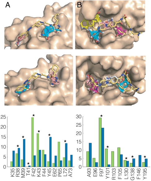
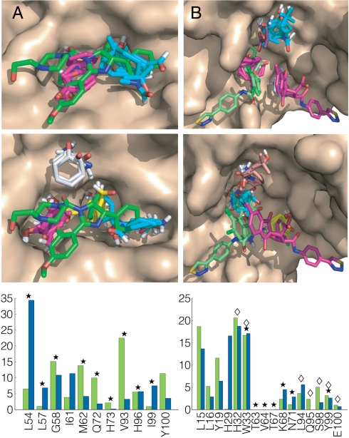
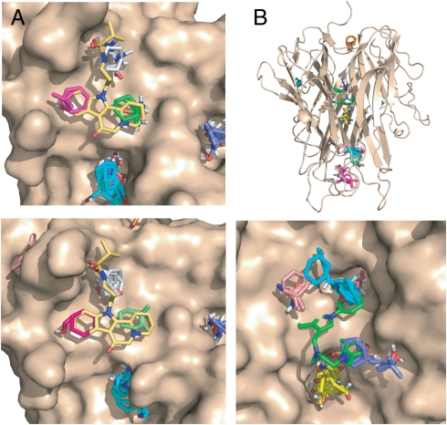
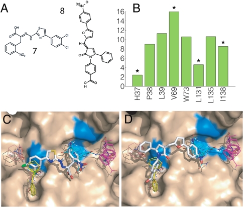
Despite the growing number of examples of small-molecule inhibitors that disrupt protein-protein interactions (PPIs), the origin of druggability of such targets is poorly understood. To identify druggable sites in protein-protein interfaces we combine computational solvent mapping, which explores the protein surface using a variety of small "probe" molecules, with a conformer generator to account for side-chain flexibility. Applications to unliganded structures of 15 PPI target proteins show that the druggable sites comprise a cluster of binding hot spots, distinguishable from other regions of the protein due to their concave topology combined with a pattern of hydrophobic and polar functionality. This combination of properties confers on the hot spots a tendency to bind organic species possessing some polar groups decorating largely hydrophobic scaffolds. Thus, druggable sites at PPI are not simply sites that are complementary to particular organic functionality, but rather possess a general tendency to bind organic compounds with a variety of structures, including key side chains of the partner protein. Results also highlight the importance of conformational adaptivity at the binding site to allow the hot spots to expand to accommodate a ligand of drug-like dimensions. The critical components of this adaptivity are largely local, involving primarily low energy side-chain motions within 6 Å of a hot spot. The structural and physicochemical signature of druggable sites at PPI interfaces is sufficiently robust to be detectable from the structure of the unliganded protein, even when substantial conformational adaptation is required for optimal ligand binding.


Conflict of interest statement

The authors declare no conflict of interest.

Figures

Fig. 1.
Fig. 1.
Mapping results for IL-2 and Bcl-xL. (A) Mapping of IL-2. (Top) Unliganded IL-2 (PDB ID code 1f47). CS1 (cyan, 20) is in the rigid hydrophilic pocket close to the site that binds the guanido group of compound 1, CS4 (salmon, 10) is at the adaptive hydrophobic pocket overlapping with the dichlorophenyl moiety. The number in parentheses following the color code indicates the number of probe clusters. Only the protein is used in the mapping; the inhibitor is shown for reference. CS1 is in the IL-2/IL-2Rα interface, and CS4 is close to it. We note that CS2 (17 probe clusters) and the small hot spot CS8 (4 clusters) are in the IL-2/IL-2Rβ interface, which makes this second interface also druggable. (Middle) IL-2 structure from the cocrystal with compound 1 (PDB ID code 1pw6). CS1 (cyan, 16) is now in the adaptive hydrophobic pocket, and CS3 (salmon, 13) identifies the rigid polar pocket. (Lower) Mapping fingerprints for IL-2, i.e., the percentages of nonbonded interactions between the probes and each amino acid residue. Green, unbound; blue, bound. Stars indicate residues interacting with compound 1 (Fig. S1). (B) Mapping of Bcl-xL. (Top) Unliganded Bcl-xL (PDB ID code 1r2d) with the modified conformations of the R100 and Y101 side chains. Both CS1 (cyan, 22) and CS3 (yellow, 16) are in the pocket which binds the distal 4-chlorophenyl ring of ABT-737 (compound 2 in Fig. S1). CS2 (magenta, 18) overlaps with the thiophenyl group of ABT-737. CS4 (salmon, 14) is in a pocket binding the piperazine and acylsulfonamide groups of ABT-737. (Middle) Bcl-xL from the cocrystal with ABT-737. CS1 (cyan, 28) overlaps with the thiophenyl, CS2 (magenta, 25) and CS4 (salmon, 9) are in the pocket binding the chlorophenyl, and CS5 (gray, 8) is in the middle. (Lower) Mapping fingerprints for Bcl-xL; green, unbound; blue, bound. Stars indicate residues interacting with ABT-737.
Fig. 2.
Fig. 2.
Mapping results for MDM2 and HPV-11 E2. (A) Mapping of MDM2. (Top) Unliganded MDM2 (PDB ID code 4z1m). CS1 (cyan, 21) is in the pocket that binds the two bromophenyl groups of compound 3 (Fig. S1), shown in green. CS2 (magenta, 21) superimposes the ethyoxyphenyl group. (Middle) MDM2 from the cocrystal with compound 3 (PDB ID code 1rv1). CS1 (cyan, 21) and CS2 (magenta, 20) identify the subsites that bind the bromophenyl and ethoxyphenyl groups of compound 3. CS3 (yellow, 19) defines a distinct subsite, which binds the second bromophenyl moiety of compound 3. CS5 (gray, 8) does not directly interact with the inhibitor. (Lower) Mapping fingerprints for MDM2; green, unbound; blue, bound. Stars indicate residues interacting with compound 3. (B) Mapping of HPV-11 E2. (Top) Unliganded HPV-11 E2 (PDB ID code 1r6k) after adjusting the “movable” side chains. CS1 (cyan, 20) is in a pocket that occupied by an isobutyric acid molecule in the inhibitor-bound structure. CS2 (magenta, 18) overlaps with the indandione moiety of inhibitor 1 (shown in green), and CS5 (gray, 10) the dichlorophenyl group of inhibitor 2 (shown in magenta). (Middle) HPV-11 E2 from the cocrystal with compound 4 (PDB ID code 1r6n). CS1 (cyan, 20) is in the pocket binding the indandione moiety of inhibitor 1 (green). CS3 (yellow, 16) overlaps with the dichlorophenyl of inhibitor 2 (magenta). CS4 (salmon, 16) is in the pocket that is occupied by an isobutyric acid. (Lower) Mapping fingerprints for HPV-11 E2; green, unbound; blue, bound. Stars and diamonds indicate residues interacting with inhibitors 1 and 2, respectively.
Fig. 3.
Fig. 3.
Mapping results for Zip and TNF-α. A. Mapping of ZipA. (Top) Unliganded ZipA (PDB ID code 1f46). CS1 (cyan, 17) is not in the ZipA/FtsZ peptide interface and does not overlap with the inhibitor. The consensus sites in the interface are CS8 (green, 6), CS9 (gray, 5), and CS11 (magenta, 3). (Lower) ZipA cocrystallized with compound 5 (Fig. S1). (PDB ID code 1s1s). The consensus sites in the interface are CS4 (gray, 10), CS8 (green, 4), and CS10 (magenta, 2). B. Mapping of TNFα. Top panel: Intact TNFα trimer. The only consensus site in the TNFα-TNFR1 interface is CS7 (orange, 5). The hot spots CS1 (cyan, 22), CS2 (magenta, 20), CS3 (19, yellow), CS4 (salmon, 7), and CS6 (blue, 6) are in the interior of the protein. (Lower) Results for the A and B chains of TNFα obtained by removing chain C from the trimeric structure (PDB ID code 1tnf). CS2 (cyan, 19) overlaps with the trifluoromethylphenyl indole moiety of compound 6 (shown in green), CS3 (yellow, 19) is close to K98 and Y119, CS4 (salmon, 10) is near Y59 and Y151, and CS6 (blue, 9) overlaps with the dimethyl chromone group.
Fig. 4.
Fig. 4.
Mapping results for eIF4E. (A) eIF4E inhibitors: 4EGI-1 (compound 7) and 4E1RCat (compound 8). (B) Mapping fingerprint for eIF4E. Stars indicate the eIF4E residues (H37, V69, L131, and I138) that, based on NMR line broadening (20), interact with 4EGI-1. (C) Mapping of the eIF4E structure from the eIF4E/4E-BP1 complex (PDB ID code 1wkw). The hot spots shown are CS1 (cyan, 24), CS2 (magenta, 22), CS3 (yellow, 19), and CS4 (salmon, 10). The inhibitor 4EGI-1 (KD ∼ 25 μM) is docked to best superimpose the hot spots. Residues V69, L131, and I138 are colored blue. H37 is on the flexible amino terminal portion of the protein and is not shown. (D) The inhibitor 4E1RCat (KD ∼ 4 μM) is docked to superimpose the hot spots.

References

    1. Wells J, McClendon C. Reaching for high-hanging fruit in drug discovery at protein-protein interfaces. Nature. 2007;450:1001–1009. - PubMed
    1. Whitty A, Kumaravel G. Between a rock and a hard place? Nat Chem Biol. 2006;2:112–118. - PubMed
    1. Berg T. Small-molecule inhibitors of protein-protein interactions. Curr Opin Drug Discov Devel. 2008;11:666–674. - PubMed
    1. Fuller JC, Burgoyne NJ, Jackson RM. Predicting druggable binding sites at the protein-protein interface. Drug Disc Today. 2009;14:155–161. - PubMed
    1. Laskowski RA, Luscombe NM, Swindells MB, Thornton JM. Protein clefts in molecular recognition and function. Protein Sci. 1996;5:2438–2452. - PMC - PubMed

Publication types

LinkOut - more resources