. 2011 Dec;10(12):M111.011213.
doi: 10.1074/mcp.M111.011213. Epub 2011 Aug 1.

The RNA-binding protein Rrm4 is essential for efficient secretion of endochitinase Cts1

Affiliations

The RNA-binding protein Rrm4 is essential for efficient secretion of endochitinase Cts1

Janine Koepke et al. Mol Cell Proteomics. 2011 Dec.

Abstract

Long-distance transport of mRNAs is crucial in determining spatio-temporal gene expression in eukaryotes. The RNA-binding protein Rrm4 constitutes a key component of microtubule-dependent mRNA transport in filaments of Ustilago maydis. Although a number of potential target mRNAs could be identified, cellular processes that depend on Rrm4-mediated transport remain largely unknown. Here, we used differential proteomics to show that ribosomal, mitochondrial, and cell wall-remodeling proteins, including the bacterial-type endochitinase Cts1, are differentially regulated in rrm4Δ filaments. In vivo UV crosslinking and immunoprecipitation and fluorescence in situ hybridization revealed that cts1 mRNA represents a direct target of Rrm4. Filaments of cts1Δ mutants aggregate in liquid culture suggesting an altered cell surface. In wild type cells Cts1 localizes predominantly at the growth cone, whereas it accumulates at both poles in rrm4Δ filaments. The endochitinase is secreted and associates most likely with the cell wall of filaments. Secretion is drastically impaired in filaments lacking Rrm4 or conventional kinesin Kin1 as well as in filaments with disrupted microtubules. Thus, Rrm4-mediated mRNA transport appears to be essential for efficient export of active Cts1, uncovering a novel molecular link between mRNA transport and the mechanism of secretion.


Figures
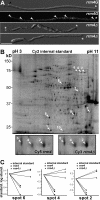
Fig. 1.
Fig. 1.
DIGE identified ten protein variants with altered amounts in the absence of Rrm4. A, rrm4Δ strains are disturbed in filamentous growth. DIC (differential interference contrast) images and fluorescence micrographs detecting Gfp of AB33rrm4G and AB33rrm4Δ filaments are shown. Black arrowheads indicate retraction septa, white arrowheads depict Rrm4-containing particles. Growth cones are marked with asterisks (size bar = 10 μm). B, Cy2 image of a representative DIGE gel showing the internal standard of membrane-associated proteins derived from AB33 and AB33rrm4Δ filaments (size marker on the left, pH range at the top). Ten protein variants exhibiting at least 2.5-fold differences in protein amounts are indicated by numbered arrowheads. Given below are enlarged Cy5 (left) and Cy3 (right) images of the same gel visualizing spots 2, 4, and 6 in protein samples from AB33 and AB33rrm4Δ filaments, respectively. C, The three panels show graphical representations of the standardized logarithmic protein abundances for spots 2, 4, and 6 obtained from three biological replicates. The internal standard (circle) is set to 0 (rectangle, wild type replicates; triangle, rrm4Δ replicates).
Fig. 2.
Fig. 2.
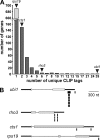
CLIP experiments reveal that cts1 mRNA is directly bound by Rrm4 in vivo. A, Bar diagram summarizing the results of high-throughput sequencing of previously generated CLIP libraries (18). Labeled arrowheads indicate the presence of relevant transcripts in a given category. B, Graphic representation of the position of unique CLIP tags in target mRNAs using the following symbols for the gene structure: exons, gray rectangles; 5′and 3′UTRs (defined as 300 nt in length), bold lines; introns, thin line. Unique CLIP tags are indicated by small filled rectangles below the gene structure (note that because of space limitations, the wide black rectangle in the 3′ UTR of ubi1 represents 28 unique CLIP tags that are only few nucleotides apart).
Fig. 3.
Fig. 3.
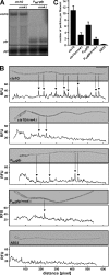
cts1G mRNA preferentially accumulates in Rrm4-dependent particles. A, Northern analysis comparing amounts of cts1G and gfp mRNA (indicated on the left) in filaments of strains AB33cts1G and AB33Ptef-gfp as well as respective rrm4Δ derivatives (indicated above). ppi mRNA encoding peptidyl-prolyl isomerase served as loading control. B, FISH analysis of AB33 filaments and derivatives (relevant alleles given on the left). Inverted fluorescence images (left) and fluorescence intensity graphs (right) are shown. In the latter, relative fluorescence signals were plotted along the longitudinal axis of the filament (x axis, distance from the rear pole). Detection of peaks (filled arrowheads) was performed using PIA. C, Bar diagram of mean particle numbers determined by PIA analysis of at least 67 filaments in three independent experiments (relevant genotypes labeled at the bottom; error bars, S.E., n = 3).
Fig. 4.
Fig. 4.
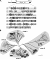
cts1 (um10419) encodes a bacterial-type chitinase of glycoside hydrolase family 18. A, Upper part, schematic drawing of Cts1 containing a Glyco_18 domain (SMART accession number SM00636) known from members of the glycoside hydrolase family 18. Amino acid positions are given above. Lower part, the sequence of the Cts1 Glyco_18 domain is aligned to sequences from fungal and bacterial chitinases CiX1 (C. immitis, 43) and ChiA (S. marcescens, 42), respectively. Identical amino acids in two or all three sequences are shaded in gray or black, respectively. Arrowheads indicate conserved amino acids which form the substrate binding pocket and the active site of CiX1 (black) and ChiA (gray) according to structural data. B, Unrooted phylogenetic tree of 80 enzymes of the glycoside hydrolase family 18. Representatives from bacteria (S. marcescens, V. fisheri, and V. harveyi; shaded in white), higher plants (N. tabacum and A. thaliana; shaded in dark gray), and fungi (ascomycetes and basidiomycetes are shaded in light gray and black, respectively) are shown. Maximum Likelihood bootstrap values (1000 replicates) are given for the main branches (asterisks indicate values above 90%). Branch lengths correspond to genetic distances. Organisms are abbreviated as follows: Sm, S. marcescens; Vf, V. fisheri; Vh, V. harveyi; Nt, N. tabacum; At, A. thaliana; Mg, M. globosa; Sc, S. cerevisiae; Sp, S. pombe; Cn, C. neoformans, Af, A. fumigatus; Hj, H. jecorina; Lb, L. bicolor; Pg, P. graminis. Accession numbers are given in Supplementary data. The three predicted endochitinases from U. maydis are indicated as UmCts1 (um10419), um06190, and um02758.
Fig. 5.
Fig. 5.
cts1Δ filaments aggregate in liquid culture. A, AB33 and derivatives were grown for 16 h in liquid minimal medium. B, Edge of colonies of strains AB33 and AB33cts1Δ incubated under filament-inducing conditions on minimal medium plates for 24 h. C, Results of plant infection experiments with solopathogenic strains SG200 (47) and SG200cts1Δ. The percentage of plants with typical disease symptoms is given. At least 110 plants were infected with each strain.
Fig. 6.
Fig. 6.
The amount of Cts1G is increased during filamentation. Western blot analysis comparing budding cells and filaments of strains AB33cts1G and AB33cts1G/rrm4Δ. Total protein extracts (A) as well as soluble (B) or membrane-associated (C) fractions were investigated. Detected proteins are given on the right (marker in kDa). Detection of α-tubulin Tub1 (in A and B) or Deep Purple (GE Healthcare) protein staining (C) served as loading controls.
Fig. 7.
Fig. 7.
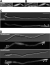
Cts1G accumulates at growth cones. DIC images and fluorescence micrographs detecting Gfp of AB33cts1G and AB33cts1G/rrm4Δ are shown. Budding cells (A and B) and filaments (C and D) were analyzed in the upper and lower part, respectively (genotypes are indicated inside DIC images, size bar = 10 μm). White arrowheads in DIC images and fluorescence micrographs indicate retraction septa and accumulations of Cts1G at poles, respectively. D, A bipolar (top) as well as a unipolar filament (bottom) of AB33cts1G/rrm4Δ are shown.
Fig. 8.
Fig. 8.
Secretion of Cts1 is impaired in rrm4Δ and kin1Δ strains. A, Bar diagram comparing endochitinolytic activity (relative fluorescence units, RFU) in budding cells and filaments (genotype is given below; error bars represent standard deviation, n = 3). B, Bar diagram comparing endochitinolytic activity of filaments of AB33 and AB33rrm4Δ in the presence of increasing concentrations of digitonin (note that fluorescence was measured with optimal gain settings, see Experimental procedures; RFU values cannot be directly compared between panels). C, Bar diagram comparing endochitinolytic activity in filaments of different AB33 derivatives (genotypes are given). Strains were either treated with solvent control or with 20 μm benomyl for 4 h. D, Bar diagram showing results from experimental setup as in (C) including additional treatment with 162 μm digitonin for membrane permeabilization as indicated below.

Similar articles

Cited by

References

    1. Gierz G., Bartnicki-Garcia S. (2001) A three-dimensional model of fungal morphogenesis based on the vesicle supply center concept. J. Theor. Biol. 208, 151–164 - PubMed
    1. Fischer R., Zekert N., Takeshita N. (2008) Polarized growth in fungi - interplay between the cytoskeleton, positional markers and membrane domains. Mol. Microbiol. 68, 813–826 - PubMed
    1. Harris S. D. (2006) Cell polarity in filamentous fungi: shaping the mold. Int. Rev. Cytol. 251, 41–77 - PubMed
    1. Steinberg G. (2007) Hyphal growth: a tale of motors, lipids, and the Spitzenkörper. Euk. Cell 6, 351–360 - PMC - PubMed
    1. Brefort T., Doehlemann G., Mendoza-Mendoza A., Reissmann S., Djamei A., Kahmann R. (2009) Ustilago maydis as a Pathogen. Annu. Rev. Phytopathol. 47, 423–445 - PubMed

Publication types

MeSH terms