. 2011 Sep 23;286(38):33652-60.
doi: 10.1074/jbc.M111.241687. Epub 2011 Aug 1.

The five near-iron transporter (NEAT) domain anthrax hemophore, IsdX2, scavenges heme from hemoglobin and transfers heme to the surface protein IsdC

Affiliations

The five near-iron transporter (NEAT) domain anthrax hemophore, IsdX2, scavenges heme from hemoglobin and transfers heme to the surface protein IsdC

Erin Sarah Honsa et al. J Biol Chem. .

Abstract

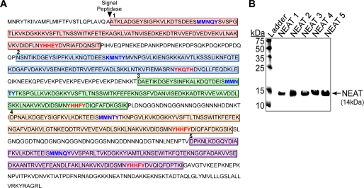
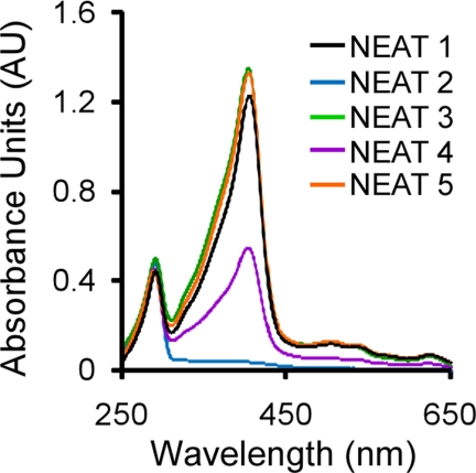
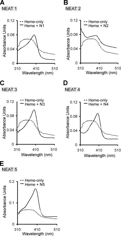
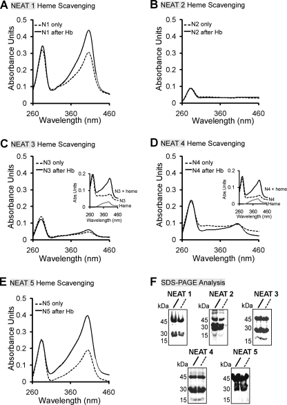
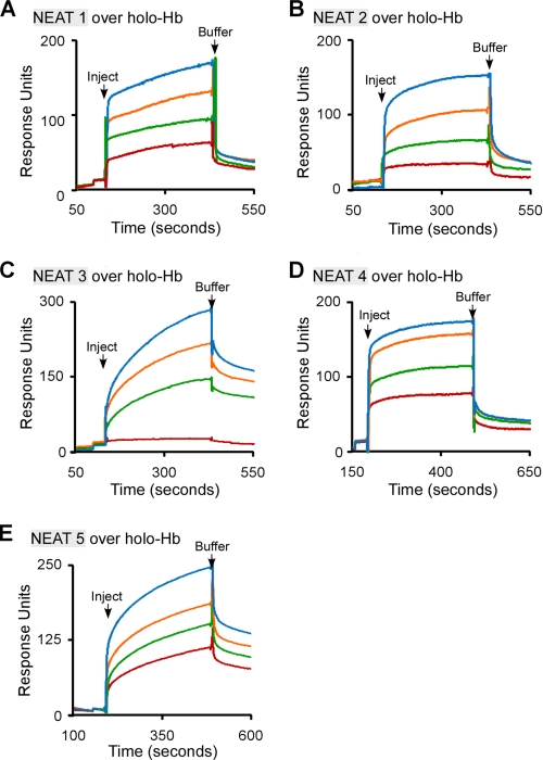
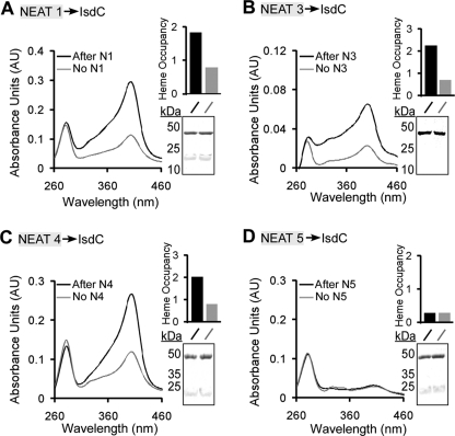
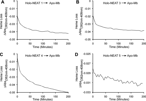
Pathogenic bacteria require iron to replicate inside mammalian hosts. Recent studies indicate that heme acquisition in Gram-positive bacteria is mediated by proteins containing one or more near-iron transporter (NEAT) domains. Bacillus anthracis is a spore-forming, Gram-positive pathogen and the causative agent of anthrax disease. The rapid, extensive, and efficient replication of B. anthracis in host tissues makes this pathogen an excellent model organism for the study of bacterial heme acquisition. B. anthracis secretes two NEAT hemophores, IsdX1 and IsdX2. IsdX1 contains a single NEAT domain, whereas IsdX2 has five, a novel property among hemophores. To understand the functional significance of harboring multiple, non-identical NEAT domains, we purified each individual NEAT domain of IsdX2 as a GST fusion and analyzed the specific function of each domain as it relates to heme acquisition and transport. NEAT domains 1, 3, 4, and 5 all bind heme, with domain 5 having the highest affinity. All NEATs associate with hemoglobin, but only NEAT1 and -5 can extract heme from hemoglobin, seemingly by a specific and active process. NEAT1, -3, and -4 transfer heme to IsdC, a cell wall-anchored anthrax NEAT protein. These results indicate that IsdX2 has all the features required to acquire heme from the host and transport heme to the bacterial cell wall. Additionally, these results suggest that IsdX2 may accelerate iron import rates by acting as a "heme sponge" that enhances B. anthracis replication in iron-starved environments.


Figures

FIGURE 1.
FIGURE 1.
In silico analysis and purification of IsdX2 NEAT domains. A, the full 885-amino acid sequence of IsdX2 is shown. The lip region (blue text) and the YXXXY signature sequence within the β8-sheet (red text) are indicated for each color-coded NEAT domain. The arrowhead indicates the predicted site of cleavage by signal peptidase. B, each NEAT protein was expressed in E. coli and purified by GST affinity chromatography. Approximately 5 μg of each thrombin-cleaved preparation was applied to SDS-PAGE and analyzed by Coomassie stain. All NEAT domains were obtained in a pure form at the correct predicted size (14 kDa).
FIGURE 2.
FIGURE 2.
Analysis of the purified IsdX2 NEAT domains. Each recombinant NEAT domain was purified as described under “Experimental Procedures.” The resulting recombinant proteins (∼15 μm) were analyzed for the presence of a Soret band at 400 nm. Specific Soret wavelengths were as follows: 405 nm for NEAT1 (black); 404 nm for NEAT3 (green) and NEAT5 (orange); 403 nm for NEAT4 (purple). NEAT2 does not absorb light at the Soret wavelength, suggesting that it cannot bind heme (blue).
FIGURE 3.
FIGURE 3.
Heme binding by the IsdX2 NEAT domains. A–E, each apo-NEAT (4.5 μm) was incubated with 2.5 μm heme for 15 min (solid lines). Spectroscopic scans of NEAT1 (A), NEAT2 (B), NEAT3 (C), NEAT4 (D), and NEAT5 (E) are shown and compared with the heme-only control reactions (2.5 μm; dotted lines).
FIGURE 4.
FIGURE 4.
Heme scavenging from Hb by the IsdX2 NEAT domains. A–E, NEAT domain 1 (12 μm; A), 2 (16.5 μm; B), 3 (15 μm; C), 4 (13.5 μm; D), or 5 (15 μm; E) was coupled to glutathione-Sepharose, and the complexes were incubated with holo-Hb (2.5 μm monomer). NEAT proteins complexed to the resin were next separated from Hb, the resin was washed, GST-NEATs were eluted, and each eluate (solid lines) was analyzed by comparing its Soret intensity with an equivalent amount of GST-NEAT protein incubated with Tris-HCl buffer instead of Hb (dotted lines; compare solid with dotted lines at 400 nm). Quantitation of the molar concentration of heme in each reaction after elution yielded the following values: NEAT1 + buffer/+Hb = 0.7 μm/2.5 μm; NEAT3 + buffer/+Hb = 0.16 μm/0.28 μm; NEAT4 + buffer/+Hb = 0.17 μm/0.22 μm; and NEAT5 + buffer/+Hb = 0.83/2.6 μm. Insets C and D, the ability of NEAT3 and -4 to bind heme was tested by incubating the NEAT-only elutions from the heme scavenging experiments with 2.5 μm heme. F, Coomassie. Eluates (10 μl) from each GST-NEAT elution were analyzed by SDS-PAGE. GST-NEATn migrates at ∼42 kDa. The bands at ∼26 and 14 kDa for each GST-NEATn elution represent free GST and NEAT, respectively, due to some proteolysis during the purification and/or experiment. Each result is a single representation of three independent experiments.
FIGURE 5.
FIGURE 5.
The association of the IsdX2 NEAT domains with holo-Hb. A–E, recombinant NEAT1 (1–20 μm; A), NEAT2 (1–15 μm; B), NEAT3 (1–17 μm; C), NEAT4 (1–14 μm; D), or NEAT5 (1–12 μm; E) was injected at a constant flow rate of 20 μl/min over 3600 RU of immobilized holo-Hb. Association and dissociation phases were monitored for 300 s by observing changes in the RUs with time. The dissociation constants (KD) were calculated assuming a 1:1 binding model (see “Experimental Procedures”) and represent the mean and S.D. values of at least three different concentrations from three independent determinations. KD was as follows: NEAT1 = 4.1 ± 1.4 × 10⁻⁸ m; NEAT2 = 4.4 ± 1.7 × 10⁻⁶ m; NEAT3 = 4.6 ± 0.8 × 10⁻⁶ m; NEAT4 = 1.8 ± 0.1 × 10⁻⁶ m; and NEAT5 = 7.5 ± 3 × 10⁻⁷. All sensorgrams analyzed had a χ2 value of <2. The S.D. values for all values were calculated from the average of two concentrations for each NEAT/IsdX2 sensorgram, from three independent experiments with multiple preparations of the same protein (n = 9).
FIGURE 6.
FIGURE 6.
Heme transfer from the IsdX2 domains to IsdC. A–D, holo-NEAT domain 1 (12 μm; A), 3 (14 μm; B), 4 (6 μm; C), or 5 (7.5 μm; D) was incubated with GST-IsdC (7 μm) that was coupled to glutathione-Sepharose resin, and complexes were washed, GST-IsdC was eluted, and eluate was analyzed by Soret spectroscopy (Fig. 6, A–D, compare black with gray lines at 400 nm). The relative heme occupancy represents the ratio of the absorbance at 400 nm to that at 280 nm (black bars, IsdC after incubation with a NEAT domain; gray bars, IsdC after incubation with a buffer control). Insets A–D, the reactions (10 μl) from each GST-IsdC elution were analyzed by SDS-PAGE. GST-IsdC migrates at ∼45 kDa, and holo-NEATs migrate at 14 kDa. The bands at 22 kDa represent free IsdC due to some proteolysis during the experiment. Each result is a single representation of three independent experiments.
FIGURE 7.
FIGURE 7.
Dissociation of heme from IsdX2 NEAT domains. A–D, holo-NEAT1 (1.17 μm; A), holo-NEAT3 (0.93 μm; B), holo-NEAT4 (1.5 μm; C), or holo-NEAT5 (0.87 μm; D) was mixed with 47 μm apo-Mb for 20 h. Spectral changes at 408 nm, relative to a control wavelength at 382 nm, were measured using a conventional spectrophotometer. The rate constants derived from these experiments are reported in Table 1.

Similar articles

Cited by

References

    1. Crosa J. H., Mey A. R., Payne S. M. (2004) Iron Transport in Bacteria, American Society of Microbiology Press, Washington, D. C
    1. Heinemann I. U., Jahn M., Jahn D. (2008) Arch. Biochem. Biophys. 474, 238–251 - PubMed
    1. De Domenico I., McVey Ward D., Kaplan J. (2008) Nat. Rev. Mol. Cell Biol. 9, 72–81 - PubMed
    1. Wandersman C., Delepelaire P. (2004) Annu. Rev. Microbiol. 58, 611–647 - PubMed
    1. Cescau S., Cwerman H., Létoffé S., Delepelaire P., Wandersman C., Biville F. (2007) Biometals 20, 603–613 - PubMed

Publication types