

Gene expression profiling suggests a pathological role of human bone marrow-derived mesenchymal stem cells in aging-related skeletal diseases
- PMID: 21808097
- PMCID: PMC3181167
- DOI: 10.18632/aging.100355
Gene expression profiling suggests a pathological role of human bone marrow-derived mesenchymal stem cells in aging-related skeletal diseases
Abstract
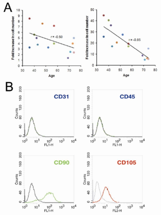
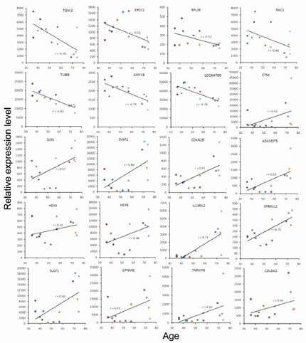
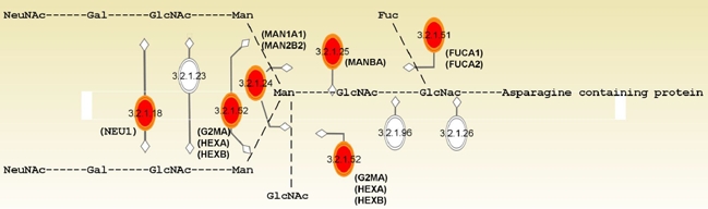
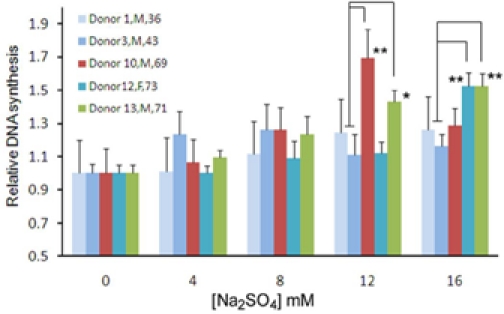
Aging is associated with bone loss and degenerative joint diseases, in which the aging of bone marrow-derived mesenchymal stem cell (bmMSC)[1] may play an important role. In this study, we analyzed the gene expression profiles of bmMSC from 14 donors between 36 and 74 years old, and obtained age-associated genes (in the background of osteoarthritis) and osteoarthritis-associated genes (in the background of old age). Pathway analysis of these genes suggests that alterations in glycobiology might play an important role in the aging of human bmMSC. On the other hand, antigen presentation and signaling of immune cells were the top pathways enriched by osteoarthritis-associated genes, suggesting that alteration in immunology of bmMSC might be involved in the pathogenesis of osteoarthritis. Most intriguingly, we found significant age-associated differential expression of HEXA, HEXB, CTSK, SULF1, ADAMTS5, SPP1, COL8A2, GPNMB, TNFAIP6, and RPL29; those genes have been implicated in the bone loss and the pathology of osteoporosis and osteoarthritis in aging. Collectively, our results suggest a pathological role of bmMSC in aging-related skeletal diseases, and suggest the possibility that alteration in the immunology of bmMSC might also play an important role in the etiology of adult-onset osteoarthritis.
Figures
Comment in
-
Altered glycobiology of stem cells linked to age-related osteoarthritis.Aging (Albany NY). 2011 Jul;3(7):663-4. doi: 10.18632/aging.100356. Aging (Albany NY). 2011. PMID: 21808096 Free PMC article.
Similar articles
-
Differential Gene Expression between Limbal Niche Progenitors and Bone Marrow Derived Mesenchymal Stem Cells.Int J Med Sci. 2020 Feb 10;17(4):549-557. doi: 10.7150/ijms.40881. eCollection 2020. Int J Med Sci. 2020. PMID: 32174786 Free PMC article.
-
Mesenchymal Stromal Cell-Derived Exosomes Affect mRNA Expression and Function of B-Lymphocytes.Front Immunol. 2018 Dec 21;9:3053. doi: 10.3389/fimmu.2018.03053. eCollection 2018. Front Immunol. 2018. PMID: 30622539 Free PMC article.
-
Comprehensive analysis of skeletal muscle- and bone-derived mesenchymal stem/stromal cells in patients with osteoarthritis and femoral neck fracture.Stem Cell Res Ther. 2020 Apr 3;11(1):146. doi: 10.1186/s13287-020-01657-z. Stem Cell Res Ther. 2020. PMID: 32245507 Free PMC article.
-
Bone marrow mesenchymal stem cells: fat on and blast off by FGF21.Int J Biochem Cell Biol. 2013 Mar;45(3):546-9. doi: 10.1016/j.biocel.2012.12.014. Epub 2012 Dec 25. Int J Biochem Cell Biol. 2013. PMID: 23270727 Free PMC article. Review.
-
Application of Cell Therapy for Anti-Aging Facial Skin.Curr Stem Cell Res Ther. 2019;14(3):244-248. doi: 10.2174/1574888X13666181113113415. Curr Stem Cell Res Ther. 2019. PMID: 30421684 Review.
Cited by
-
Glycoprotein nonmetastatic melanoma protein B regulates lysosomal integrity and lifespan of senescent cells.Sci Rep. 2022 Apr 20;12(1):6522. doi: 10.1038/s41598-022-10522-3. Sci Rep. 2022. PMID: 35444208 Free PMC article.
-
Korean mistletoe lectin regulates self-renewal of placenta-derived mesenchymal stem cells via autophagic mechanisms.Cell Prolif. 2012 Oct;45(5):420-9. doi: 10.1111/j.1365-2184.2012.00839.x. Cell Prolif. 2012. PMID: 22925501 Free PMC article.
-
Zinc finger protein ZFP36L1 promotes osteoblastic differentiation but represses adipogenic differentiation of mouse multipotent cells.Oncotarget. 2017 Mar 28;8(13):20588-20601. doi: 10.18632/oncotarget.15246. Oncotarget. 2017. PMID: 28206953 Free PMC article.
-
Zinc finger factor 521 enhances adipogenic differentiation of mouse multipotent cells and human bone marrow mesenchymal stem cells.Oncotarget. 2015 Jun 20;6(17):14874-84. doi: 10.18632/oncotarget.3900. Oncotarget. 2015. PMID: 26008984 Free PMC article.
-
MSC-sEVs exacerbate senescence by transferring bisecting GlcNAcylated GPNMB.Stem Cell Res Ther. 2025 Jan 23;16(1):23. doi: 10.1186/s13287-025-04140-9. Stem Cell Res Ther. 2025. PMID: 39849576 Free PMC article.
References
-
- Friedenstein AJ. Precursor cells of mechanocytes. Int Rev Cytol. 1976;47:327–359. - PubMed
-
- Yoo JU, Johnstone B. The role of osteochondral progenitor cells in fracture repair. Clin Orthop Relat Res. 1998:S73–81. - PubMed
-
- Quarto R, Thomas D, Liang CT. Bone progenitor cell deficits and the age-associated decline in bone repair capacity. Calcif Tissue Int. 1995;56:123–129. - PubMed
-
- Tanaka H, Liang CT. Effect of platelet-derived growth factor on DNA synthesis and gene expression in bone marrow stromal cells derived from adult and old rats. J Cell Physiol. 1995;164:367–375. - PubMed
-
- Tanaka H, Liang CT. Mitogenic activity but not phenotype expression of rat osteoprogenitor cells in response to IGF-I is impaired in aged rats. Mech Ageing Dev. 1996;92:1–10. - PubMed
Publication types
MeSH terms
Substances
LinkOut - more resources
Full Text Sources
Other Literature Sources
Medical
Molecular Biology Databases
Research Materials
Miscellaneous
