

Case Reports
doi: 10.4103/0972-2327.82803.
Hemichorea-hemiballism syndrome: A look through susceptibility weighted imaging
Affiliations
- PMID: 21808477
- PMCID: PMC3141477
- DOI: 10.4103/0972-2327.82803
Item in Clipboard
Case Reports
Hemichorea-hemiballism syndrome: A look through susceptibility weighted imaging
Ann Indian Acad Neurol.
2011 Apr.
Abstract
Hemichorea-hemiballism syndrome (HCHB) is a relatively rare cause of unilateral chorea in diabetic patients and is due to non ketotoic hyperglycaemia. Characteristic magnetic resonance (MR) findings include T1 hyperintensity in the contralateral putamen without any significant signal alteration on other conventional MR sequences. We report susceptibility weighted imaging (SWI) findings in a case of HCHB syndrome.
Keywords: Diabetes; hemichorea-hemiballism; putamen; susceptibility.
Conflict of interest statement
Figures
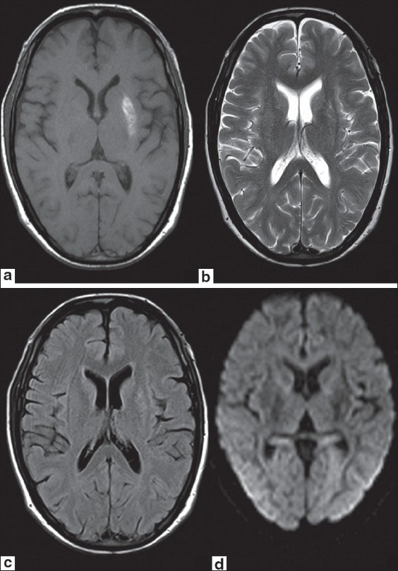
Axial T1W MR image (a) shows hyperintense signal in left lentiform nucleus. No significant signal abnormality is seen on axial T2W (b), FLAIR (c) or DW (d) images
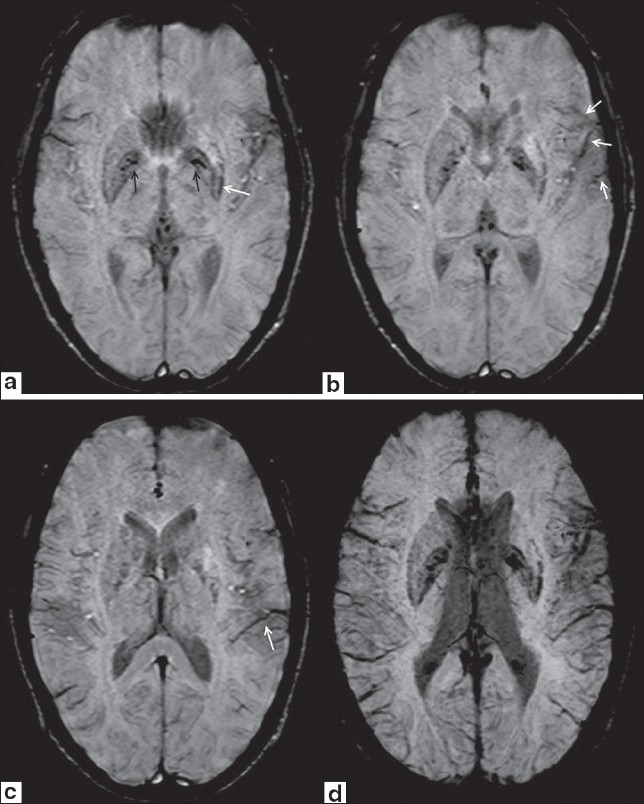
Axial SWI images show hyperintense signal in left lentiform nucleus. Symmetrical hypointensities are seen in bilateral globus palidii suggesting age-related mineralization (black arrows in 2a). Asymmetrical hypointensity is also seen in posterolateral aspect of left putamen indicating mineral deposition (white arrow in 2a). Prominent vessels appearing hypointense of SWI are seen along left sylvian fissure (white arrows in 2b) and in left temporal region (white arrow in 2c). Axial minimum intensity projection (MinIP) SWI image (2d) again shows prominent hypointense vessels along left sylvian fissure and in left temporal region to better advantage
References
-
- Cherian A, Thomas B, Baheti NN, Chemmanam T, Kesavadas C. Concepts and controversies in nonketotic hyperglycemia-induced hemichorea: Further evidence from susceptibility-weighted MR imaging. J Magn Reson Imaging. 2009;29:699–703. - PubMed
-
- Felicio AC, Chang CV, Godeiro-Junior C, Okoshi MP, Ferraz HB. Hemichorea-hemiballism as the first presentation of type 2 diabetes mellitus. Arq Neuropsiquiatr. 2008;66:249–50. - PubMed
