

Cell therapy in bone healing disorders
- PMID: 21808710
- PMCID: PMC3143975
- DOI: 10.4081/or.2010.e20
Cell therapy in bone healing disorders
Abstract
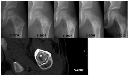
In addition to osteosynthetic stabilizing techniques and autologous bone transplantations, so-called orthobiologics play an increasing role in the treatment of bone healing disorders. Besides the use of various growth factors, more and more new data suggest that cell-based therapies promote local bone regeneration. For ethical and biological reasons, clinical application of progenitor cells on the musculoskeletal system is limited to autologous, postpartum stem cells. Intraoperative one-step treatment with autologous progenitor cells, in particular, delivered promising results in preliminary clinical studies. This article provides an overview of the rationale for, and characteristics of the clinical application of cell-based therapy to treat osseous defects based on a review of existing literature and our own experience with more than 100 patients. Most clinical trials report successful bone regeneration after the application of mixed cell populations from bone marrow. The autologous application of human bone marrow cells which are not expanded ex vivo has medico-legal advantages. However, there is a lack of prospective randomized studies including controls for cell therapy for bone defects. Autologous bone marrow cell therapy seems to be a promising treatment option which may reduce the amount of bone grafting in future.
Keywords: bone defect; cell therapy; osteoblast.; stem cell.
Figures
Similar articles
-
[Cell therapy in bone-healing disorders].Orthopade. 2010 Apr;39(4):449-62; quiz 463. doi: 10.1007/s00132-009-1583-7. Orthopade. 2010. PMID: 20182700 Review. German.
-
Osteogenic protein-1 for long bone nonunion: an evidence-based analysis.Ont Health Technol Assess Ser. 2005;5(6):1-57. Epub 2005 Apr 1. Ont Health Technol Assess Ser. 2005. PMID: 23074475 Free PMC article.
-
Guided bone regeneration in pig calvarial bone defects using autologous mesenchymal stem/progenitor cells - a comparison of different tissue sources.J Craniomaxillofac Surg. 2012 Jun;40(4):310-20. doi: 10.1016/j.jcms.2011.05.004. Epub 2011 Jun 30. J Craniomaxillofac Surg. 2012. PMID: 21723141
-
Bone marrow concentrate for autologous transplantation in minipigs. Characterization and osteogenic potential of mesenchymal stem cells.Vet Comp Orthop Traumatol. 2013;26(1):34-41. doi: 10.3415/VCOT-11-11-0165. Epub 2012 Nov 21. Vet Comp Orthop Traumatol. 2013. PMID: 23171924
-
Bone Marrow Concentrate (BMC) Therapy in Musculoskeletal Disorders: Evidence-Based Policy Position Statement of American Society of Interventional Pain Physicians (ASIPP).Pain Physician. 2020 Mar;23(2):E85-E131. Pain Physician. 2020. PMID: 32214287 Review.
Cited by
-
Cell based therapy in Parkinsonism.Transl Neurodegener. 2013 Jun 4;2(1):13. doi: 10.1186/2047-9158-2-13. Transl Neurodegener. 2013. PMID: 23734727 Free PMC article.
-
The Effect of Amniotic Tissue on Spinal Interventions: A Systematic Review.Int J Spine Surg. 2023 Feb;17(1):32-42. doi: 10.14444/8380. Epub 2022 Oct 17. Int J Spine Surg. 2023. PMID: 36253081 Free PMC article.
-
Caffeic Acid Phenethyl Ester With Mesenchymal Stem Cells Improves Behavioral and Histopathological Changes in the Rat Model of Parkinson Disease.Basic Clin Neurosci. 2022 Sep-Oct;13(5):637-646. doi: 10.32598/bcn.2021.1398.1. Epub 2022 Sep 1. Basic Clin Neurosci. 2022. PMID: 37313025 Free PMC article.
-
New bio-ceramization processes applied to vegetable hierarchical structures for bone regeneration: an experimental model in sheep.Tissue Eng Part A. 2014 Feb;20(3-4):763-73. doi: 10.1089/ten.TEA.2013.0108. Epub 2013 Dec 11. Tissue Eng Part A. 2014. PMID: 24099033 Free PMC article.
-
Sartorius muscle pedicle iliac bone graft for the treatment of avascular necrosis of femur head.J Hip Preserv Surg. 2016 Apr 25;3(3):215-22. doi: 10.1093/jhps/hnw012. eCollection 2016 Aug. J Hip Preserv Surg. 2016. PMID: 27583161 Free PMC article.
References
-
- Coipeau P, Rosset P, Langonne A, et al. Impaired differentiation potential of human trabecular bone mesenchymal stromal cells from elderly patients. Cytotherapy. 2009;11:584–94. - PubMed
-
- El Tamer MK, Reis RL. Progenitor and stem cells for bone and cartilage regeneration. J Tissue Eng Regen Med. 2009;3:327–37. - PubMed
-
- Mackay DL, Tesar PJ, Liang LN, Haynesworth SE. Characterizing medullary and human mesenchymal stem cell-derived adipocytes. J Cell Physiol. 2006;207:722–8. - PubMed
LinkOut - more resources
Full Text Sources