

Bony island within the articular cartilage of the knee in a child: a rare condition for early osteoarthritis
- PMID: 21808719
- PMCID: PMC3144001
- DOI: 10.4081/or.2011.e7
Bony island within the articular cartilage of the knee in a child: a rare condition for early osteoarthritis
Abstract
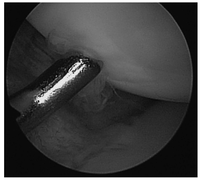
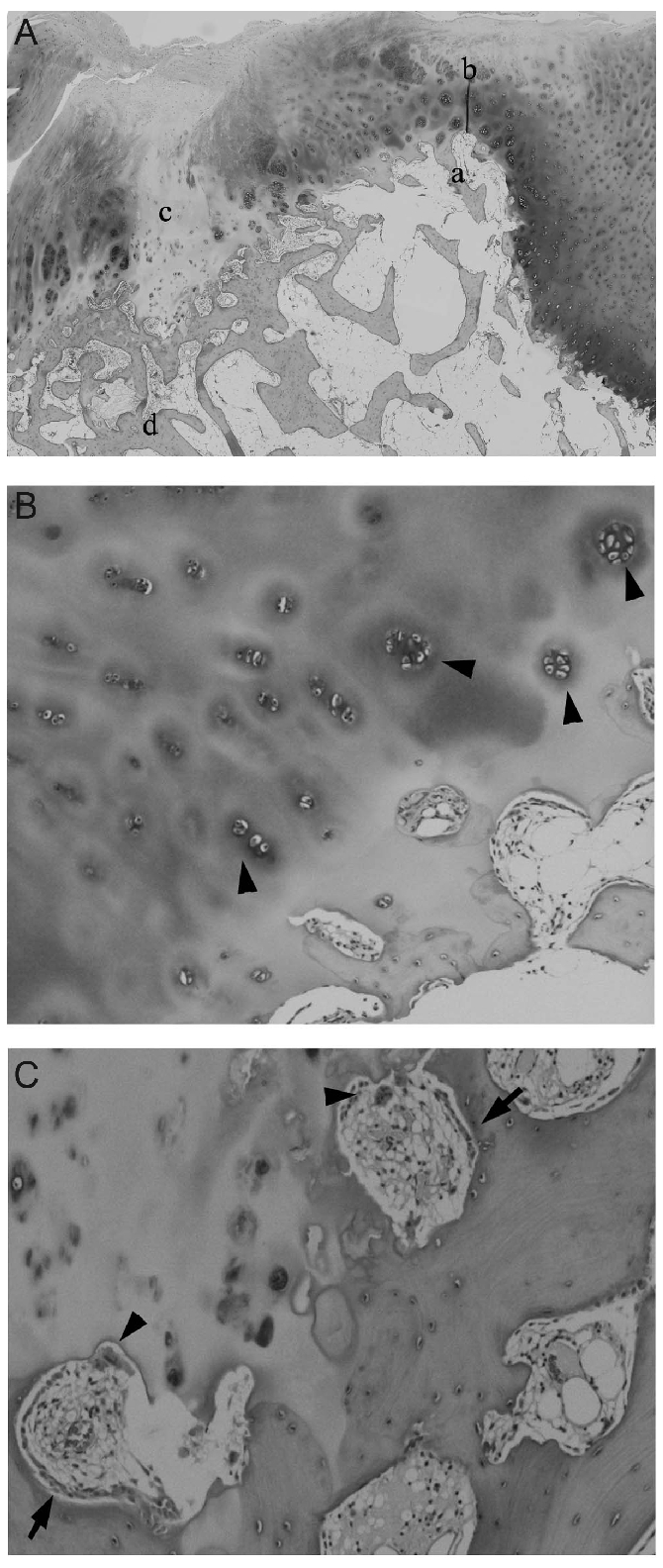
Articular cartilage is a specific type of connective tissue composed of hydrated proteoglycans within a matrix of collagen fibrils. In the elderly population, it shows degenerative changes that may results in osteoarthritis. The more severe form of osteoarthritis occasionally demonstrates bone formation within the cartilage, which is designated as a bony protuberance, however, such lesions are rare in children. This report presents the case of a 10-year-old boy with a bony protuberance within the articular cartilage of the knee. The patient initially complained of knee pain and he subsequently developed flexion contracture. Radiological and arthroscopic examinations revealed a bony protuberance in the articular cartilage and degenerative changes of the cartilage above it. He was successfully treated by the removal of the bony protuberance and osteochondral grafting. The bony protuberance may have caused cartilage degradation since the thickness of the cartilage above it was thinner than that around the lesion. The bony protuberance within the articular cartilage formed in the younger population may be a possible cause of osteoarthritis. This case is a noteworthy with regard to the pathogenesis of osteoarthritis.
Keywords: articular cartilage; bone protuberance; child.; osteoarthritis.
Conflict of interest statement
Conflict of interest: the authors report no conflicts of interest.
Figures


References
-
- Clarke HD, Scott WN, Insall JN, et al. Anatomy. In: Scott WN, editor. Surgery of the knee. 4th ed. New York: Churchill Livingstone; 2006. pp. 3–66.
-
- Buckwalter JA, Amendola A, Clark CR. Articular cartilage and meniscus: biology, biomechanics, and healing response. In: Scott WN, editor. Surgery of the knee. 4th ed. New York: Churchill Livingstone; 2006. pp. 307–16.
-
- Bergman AG, Willen HK, Lindstrand AL, Pettersson HTA. Osteoarthritis of the knee: Correlation of subchondral MR signal abnormalities with histopathologic and radiographic features. Skeletal Radiol. 1994;23:445–8. - PubMed
-
- Sugita T, Chiba T, Kawamata T, et al. Assessment of articular cartilage of the lateral tibial plateau in varus osteoarthritis of the knee. The Knee. 2000;7:217–20. - PubMed
-
- Hefti F, Krauspe R, Möller-Madsen B, et al. Osteochindritis dissecans: a multicenter study of the European Pediatric Orthopedic Society. J Pediatr Orthop B. 1999;8:231–45. - PubMed
Publication types
LinkOut - more resources
Full Text Sources