. 2012 May 1;72(6):677-89.
doi: 10.1002/pros.21471. Epub 2011 Aug 1.

The H6D variant of NAG-1/GDF15 inhibits prostate xenograft growth in vivo

Affiliations

The H6D variant of NAG-1/GDF15 inhibits prostate xenograft growth in vivo

Xingya Wang et al. Prostate. .

Abstract

Background: Non-steroidal anti-inflammatory drug-activated gene (NAG-1), a divergent member of the transforming growth factor-beta superfamily, has been implicated in many cellular processes, including inflammation, early bone formation, apoptosis, and tumorigenesis. Recent clinical studies suggests that a C to G single nucleotide polymorphism at position 6 (histidine to aspartic acid substitution, or H6D) of the NAG-1 protein is associated with lower human prostate cancer incidence. The objective of the current study is to investigate the activity of NAG-1 H6D variant in prostate cancer tumorigenesis in vivo.

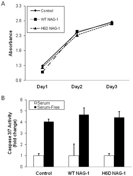
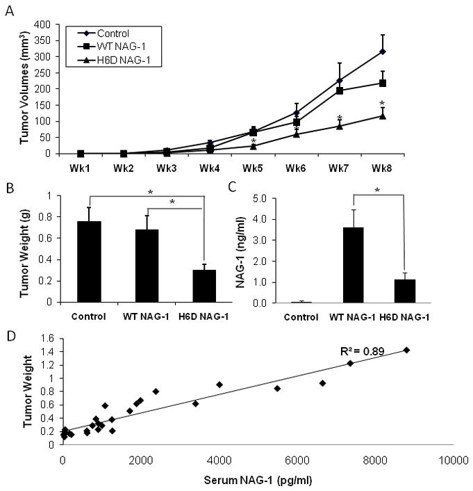
Methods: Human prostate cancer DU145 cells expressing the H6D NAG-1 or wild-type (WT) NAG-1 were injected subcutaneously into nude mice and tumor growth was monitored. Serum and tumor samples were collected for subsequent analysis.

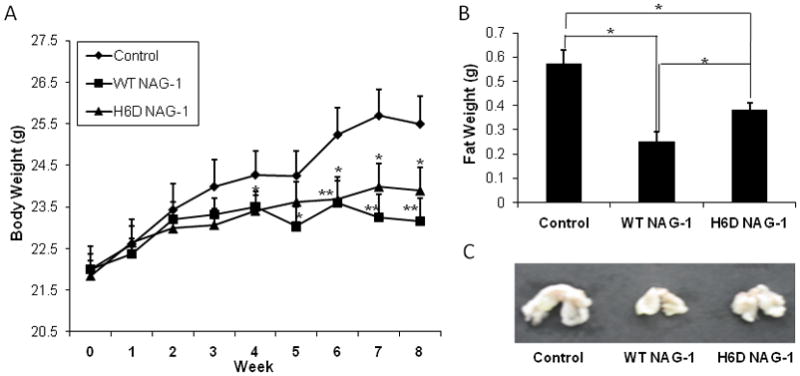
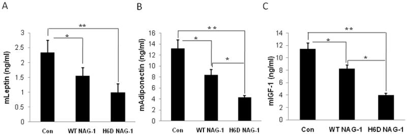
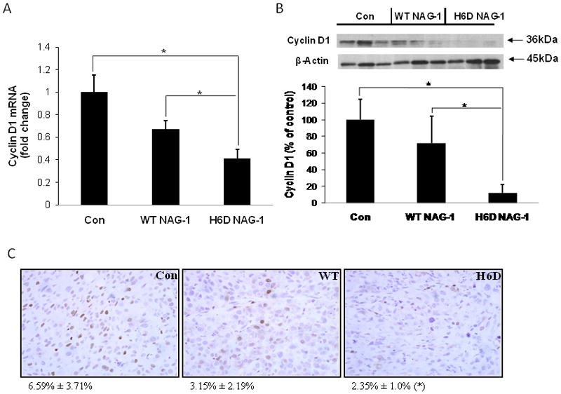
Results: The H6D variant was more potent than the WT NAG-1 and inhibited tumor growth significantly compared to control mice. Mice with tumors expressing the WT NAG-1 have greater reduced both body weight and abdominal fat than mice with H6D variant tumors suggesting different activities of the WT NAG-1 and the H6D NAG-1. A significant reduction in adiponectin, leptin, and IGF-1 serum levels was observed in the tumor-bearing mice with a more profound reduction observed with expression of H6D variant. Cyclin D1 expression was suppressed in the tumors with a dramatic reduction observed in the tumor expressing the H6D variant.

Conclusion: Our data suggest that the H6D variant of NAG-1 inhibits prostate tumorigenesis by suppressing IGF-1 and cyclin D1 expression but likely additional mechanisms are operative.


Figures

Figure 1
Figure 1. There is no difference in proliferation and apoptosis potential between the three cell lines
A: SRB proliferation assay demonstrates that the three cell lines, upon equal initial number of cells, had similar rates of proliferation. Cells (1×10⁴/well) were incubated for 24, 48, or 72 h for proliferation measurement (n=3). B: Caspase 3/7 enzyme activity assay demonstrates no difference of apoptosis between the three cell lines (n=3).
Figure 2
Figure 2. The H6D variant of NAG-1/GDF15 significantly inhibited DU145 xenograft tumor growth in vivo
A: Xenograft tumor growth measured by volume. DU145 cells (3×10⁶) carrying control vector, wild type GDF15/NAG-1, or the H6D variant were injected s.c. into nude mice (n=15 per group). Tumor growth was recorded by measuring the dimensions of tumors and presented as volume (mm3) every week for 8 wks. * p<0.05. B: Tumor weight (g) at necropsy. * p<0.01. C: Serum level of GDF15/NAG-1 (ng/ml). At necropsy, serum was collected and the circulating serum level of NAG-1/GDF15 was measured by ELISA. Data in control samples represent the background value. * p<0.01. All data are presented as mean ± SE (standard error) (n=15). D: Linear regression model indicates that the serum level of NAG-1/GDF15 (wild type or H6D) correlates with tumor sizes in WT NAG-1 and H6D NAG-1 mice. R2=0.89.
Figure 3
Figure 3. The wild type GDF15/NAG-1 most significantly reduced body weight and abdominal fat weight of the nude mice
A: Body weight (g) changes of mice during experiment were measured for 8 wks. * p<0.05; ** p<0.01. B: Weight of abdominal fat (g) was measured at necropsy. * p<0.05; ** p<0.01. C, Representative image of the mean weight of abdominal fat from each group. All data are presented as mean ± SE (n=15).
Figure 4
Figure 4. The serum level of mLeptin, mAdiponectin, and mIGF-1 were most significantly reduced in H6D of NAG-1 group
At necropsy, serum samples were collected and subjected to ELISA for the analysis of serum level of mLeptin, mAdiponectin, and mIGF-1. All data are presented as mean ± SE (n=15). * p<0.05; ** p<0.01.
Figure 5
Figure 5. The expression of Cyclin D1 was inhibited in xenograft samples from the H6D of GDF15/NAG-1 mice
A: mRNA level of Cyclin D1 in the xenograft tumors. Total RNA was extracted and Cyclin D1 expression was analyzed by qRT-PCR. Beta-Actin was used as control. All data are presented as SE ± SD (n=15). * p<0.05. B: Western blot of Cyclin D1 expression in the xenograft tumors. Total protein was extracted from three randomly selected xenograft tumor samples of each experimental group. Cell lysates were examined for Cyclin D1 expression by western blot analysis. Beta-Actin was used for loading control. Relative expression level of Cyclin D1 was analyzed by densitometric method. All data are presented as mean ± SD (n=3). * p<0.05. C: Immunohistochemical staining of Cyclin D1 in xenograft tumor samples (40×). The quantification of Cyclin D1 staining was analyzed by Image Analysis Core at NIEHS using image analysis techniques. Data is represented as % positive nuclei staining ± SD (n=6). * p<0.05 compared to control.
Figure 6
Figure 6. Model of WT Human NAG-1 (GDF15) (blue) compared with H6D mutant Human NAG-1(red)
The modeled NAG-1structure is based largely on the solved crystal structure of BMP6 (PDB: 2QCW,2R53) with which NAG-1 shares 34% sequence identity and 53% sequence similarity, also based on crystal structures of BMP7 (33% sequence identity; 52% similarity) and GDF5 (28% identity; 52% similarity). The difference in size between the N-terminal basic positively charged histidine residue in the wildtype is contrasted with the Smaller, acidic negatively charged mutant aspartic acid.

Similar articles

Cited by

References

    1. Cancer Facts and Figures. 2010.
    1. De Marzo AM, Platz EA, Sutcliffe S, Xu J, Gronberg H, Drake CG, Nakai Y, Isaacs WB, Nelson WG. Inflammation in prostate carcinogenesis. Nature reviews. 2007;7(4):256–269. - PMC - PubMed
    1. Piek E, Heldin CH, Ten Dijke P. Specificity, diversity, and regulation in TGF-beta superfamily signaling. Faseb J. 1999;13(15):2105–2124. - PubMed
    1. Eling TE, Baek SJ, Shim M, Lee CH. NSAID activated gene (NAG-1), a modulator of tumorigenesis. J Biochem Mol Biol. 2006;39(6):649–655. - PubMed
    1. Bauskin AR, Zhang HP, Fairlie WD, He XY, Russell PK, Moore AG, Brown DA, Stanley KK, Breit SN. The propeptide of macrophage inhibitory cytokine (MIC-1), a TGF-beta superfamily member, acts as a quality control determinant for correctly folded MIC-1. The EMBO journal. 2000;19(10):2212–2220. - PMC - PubMed

Publication types

MeSH terms