Comparative Study
. 2011 Oct;54(4):1282-92.
doi: 10.1002/hep.24492. Epub 2011 Aug 1.

A protective antiarrhythmic role of ursodeoxycholic acid in an in vitro rat model of the cholestatic fetal heart

Affiliations
Comparative Study

A protective antiarrhythmic role of ursodeoxycholic acid in an in vitro rat model of the cholestatic fetal heart

Michele Miragoli et al. Hepatology. 2011 Oct.

Abstract

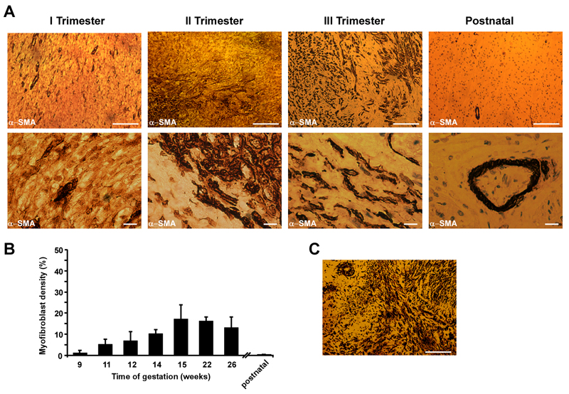
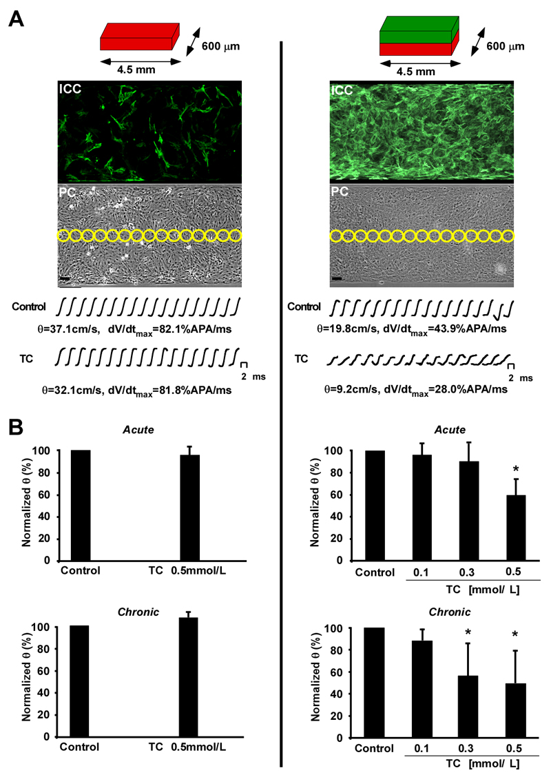
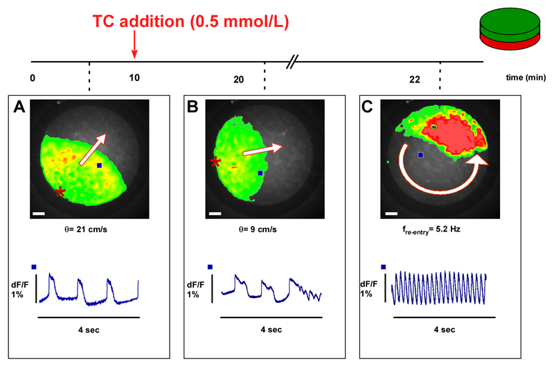
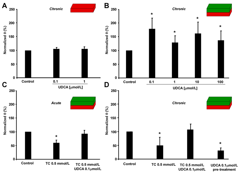
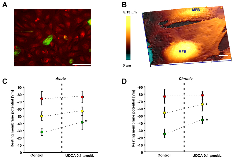
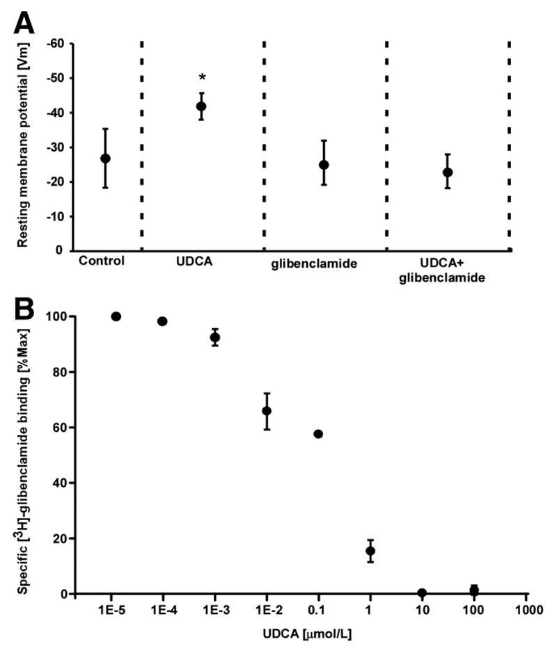
Intrahepatic cholestasis of pregnancy may be complicated by fetal arrhythmia, fetal hypoxia, preterm labor, and, in severe cases, intrauterine death. The precise etiology of fetal death is not known. However, taurocholate has been demonstrated to cause arrhythmia and abnormal calcium dynamics in cardiomyocytes. To identify the underlying reason for increased susceptibility of fetal cardiomyocytes to arrhythmia, we studied myofibroblasts (MFBs), which appear during structural remodeling of the adult diseased heart. In vitro, they depolarize rat cardiomyocytes via heterocellular gap junctional coupling. Recently, it has been hypothesized that ventricular MFBs might appear in the developing human heart, triggered by physiological fetal hypoxia. However, their presence in the fetal heart (FH) and their proarrhythmogenic effects have not been systematically characterized. Immunohistochemistry demonstrated that ventricular MFBs transiently appear in the human FH during gestation. We established two in vitro models of the maternal heart (MH) and FH, both exposed to increasing doses of taurocholate. The MH model consisted of confluent strands of rat cardiomyocytes, whereas for the FH model, we added cardiac MFBs on top of cardiomyocytes. Taurocholate in the FH model, but not in the MH model, slowed conduction velocity from 19 to 9 cm/s, induced early after depolarizations, and resulted in sustained re-entrant arrhythmias. These arrhythmic events were prevented by ursodeoxycholic acid, which hyperpolarized MFB membrane potential by modulating potassium conductance.

Conclusion: These results illustrate that the appearance of MFBs in the FH may contribute to arrhythmias. The above-described mechanism represents a new therapeutic approach for cardiac arrhythmias at the level of MFB.


Conflict of interest statement

Conflict of Interest: None

Figures

Figure 1
Figure 1
Immunohistochemical identification of cardiac myofibroblasts in human fetal ventricular tissue slices taken during the three trimesters of gestation. A. Top. Human fetal ventricular myofibroblasts react positively to α-SMA staining, showing scattered myofibroblasts areas (brown) in the second trimester, which becomes denser in the third trimester of gestation. As expected, the postnatal human heart does not contain α-SMA positive cells. Bar=100μm. Bottom. High magnification pictures depict distinct interstitial myofibroblasts (brown) in contact with resident ventricular cardiomyocytes. In postnatal samples, the α-SMA positivity is related to smooth-muscle cells lining the blood vessels. Bar=10μm. B. Transient increment of myofibroblast density throughout gestation. Myofibroblast density was analyzed from fixed ventricular preparations derived from a single fetal heart corresponding to each week of gestation, (n=10 random locations). Data: means±SD. C. Staining of infarcted human heart 6 weeks-old granulation tissue. Positive staining for α-SMA (brown) indicates presence of abundant myofibroblasts. Bar=100μm.
Figure 2
Figure 2
Optical recording of impulse propagation along homo- and heterocellular cell strands. A. Left column. Schematic representation of the maternal heart model (MH, red) consisting of strands of neonatal rat cardiomyocytes (0.6 × 4.5 mm) with little myofibroblast contamination as illustrated by the anti α-SMA staining in the immunocytochemistry (ICC) picture. Yellow circles in the corresponding phase contrast image (PC) indicate the positions of photodetectors used to record electrical activity. Bar=50 μm. Action potential upstrokes recorded during propagated activity were monophasic and fast rising and remained virtually unchanged during exposure to 0.5 mmol/L taurocholic acid (TC). B. Left column. Neither acute (top) nor chronic (bottom) application of 0.5 mmol/L TC had any significant effects on conduction velocity (θ). A. Right column. Schematic representation of the fetal heart model (FH) having identical dimensions to the maternal heart (MH) but consisting of strands of neonatal rat cardiomyocytes (red) coated with myofibroblasts (green). As indicated by the anti α-SMA staining, myofibroblasts formed a uniform cellular coat. Under control conditions, θ was slow in FH compared to the MH and acute application of 0.5 mmol/L TC led to a substantial reduction of θ and dV/dtmax. B. Right column. Both acute (top) and chronic (bottom) application of TC ranging from 0.1 to 0.5 mmol/L reduces θ in a dose dependent manner. Data: means±SD (n=9).
Figure 3
Figure 3
Example of the arrhythmia induced by taurocholic acid (TC) in a preparation consisting of a monolayer of CMs coated with a monolayer of MFBs. A. Optical recording of spontaneous electrical activity (45 bpm, bottom) originates from the periphery and propagates uniformly at 21 cm/s. B. After acute exposure to 0.5 mmol/L, TC conduction velocity (θ) is reduced from 21 to 9 cm/s. Moreover, propagated APs display early afterdepolarizations where the last activation is followed by (C) self-sustained re-entrant excitation. Frequency of rotation is 5.2 Hz. Bar=1 mm. Blue squares in the overview indicate the locations of recorded traces. Red stars indicate the origins of spontaneous electrical activation.
Figure 4
Figure 4
Effect of exposure to ursodeoxycholic acid (UDCA) on impulse propagation in the maternal (MH) and fetal heart (FH) models. A. Chronic exposure of the MH model to UDCA does not affect conduction velocity (θ, n=7). B. In contrast, chronic exposure of the FH model to increasing concentrations of UDCA significantly increases θ (n=24). C. Acute exposure of FH models to 0.5 mmol/L taurocholic acid (TC) plus 0.1 μmol/L UDCA inhibits slow conduction induced by TC alone (n=7). D. Same as C, but following chronic exposure of FH models to TC and UDCA. Again, UDCA plus TC abolishes slow conduction induced by TC alone. UDCA pre-treatment for 12 hrs (UDCA withdrawn before adding 0.5 mmol/L TC for 12 hrs) shows no cardio-protective effect (n=7). Data: mean±SD.
Figure 5
Figure 5
Effect of ursodeoxycholic acid (UDCA) on membrane potential (Vm) in the maternal heart model (MH), fetal heart model (FH) and myofibroblast monolayers. A. Myofibroblast monolayer stained for desmin (green) and vimentin (red) indicates a low contamination by desmin positive cardiomyocytes. Bar=100 μm. B. Scanning ion conductance microscopy identifies the most prominent area for impalement as regions above nuclei in the myofibroblast monolayer. Bar=10 μm. C. Acute exposure to 0.1 μmol/L UDCA significantly increases Vm only in pure myofibroblast monolayers (green dots) but not in the MH model (red dots) and FH model (yellow dots). D. Same as C for chronic exposure to UDCA. 0.1 μmol/L. UDCA significantly increases Vm in the FH model and myofibroblast monolayers but has no effect on MH model preparations. Data: means±SD, n=12.
Figure 6
Figure 6
Ursodeoxycholic acid (UDCA) hyperpolarizes myofibroblasts by increasing potassium conductance. A. Glibenclamide inhibits the effect of UDCA, indicating that UDCA is involved in the modulation of potassium conductance. Data: means±SD, n=12. B. Specific binding of [3H]-glibenclamide to sulphonylureas receptors in myofibroblast membranes is competitively displaced by increasing concentrations of UDCA. Data: means±SE n=3.

Similar articles

Cited by

References

    1. Geenes V, Williamson C. Intrahepatic cholestasis of pregnancy. World J Gastroenterol. 2009;15:2049–2066. - PMC - PubMed
    1. Glantz A, Marschall HU, Lammert F, Mattsson LA. Intrahepatic cholestasis of pregnancy: a randomized controlled trial comparing dexamethasone and ursodeoxycholic acid. Hepatology. 2005;42:1399–1405. - PubMed
    1. Al Inizi S, Gupta R, Gale A. Fetal tachyarrhythmia with atrial flutter in obstetric cholestasis. Int J Gynaecol Obstet. 2006;93:53–54. - PubMed
    1. Shand AW, Dickinson JE, D'Orsogna L. Refractory fetal supraventricular tachycardia and obstetric cholestasis. Fetal Diagn Ther. 2008;24:277–281. - PubMed
    1. Fisk NM, Storey GN. Fetal outcome in obstetric cholestasis. Br J Obstet Gynaecol. 1988;95:1137–1143. - PubMed

Publication types

MeSH terms

Substances