. 2011 Oct;29(10):1601-10.
doi: 10.1002/stem.703.

Tumor necrosis factor inhibits mesenchymal stem cell differentiation into osteoblasts via the ubiquitin E3 ligase Wwp1

Affiliations

Tumor necrosis factor inhibits mesenchymal stem cell differentiation into osteoblasts via the ubiquitin E3 ligase Wwp1

Lan Zhao et al. Stem Cells. 2011 Oct.

Abstract

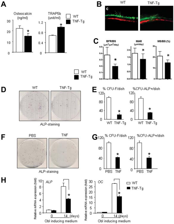
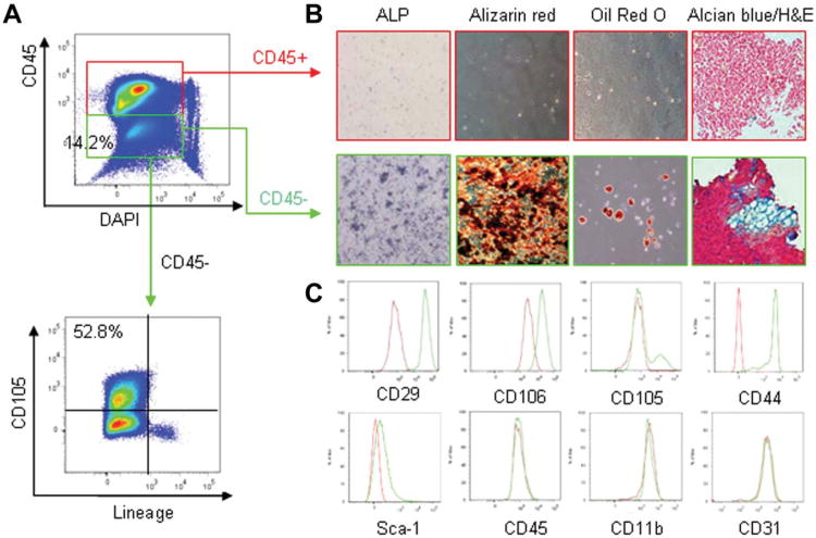
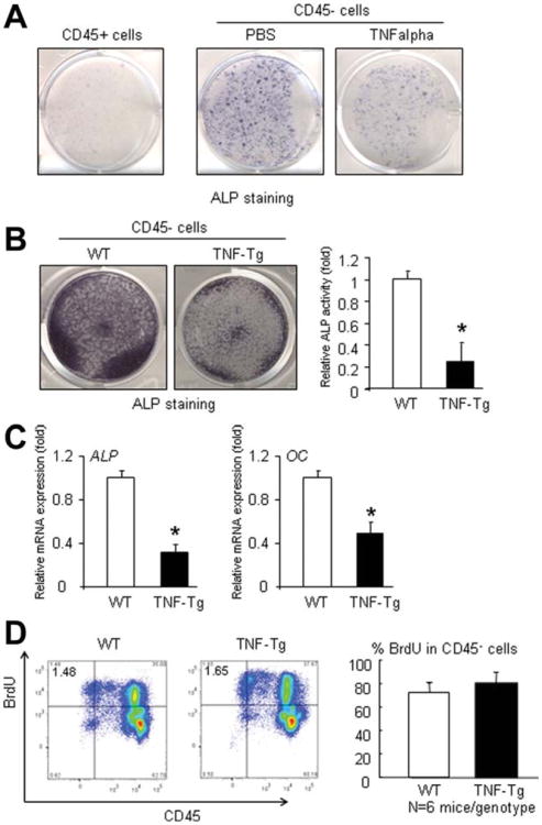
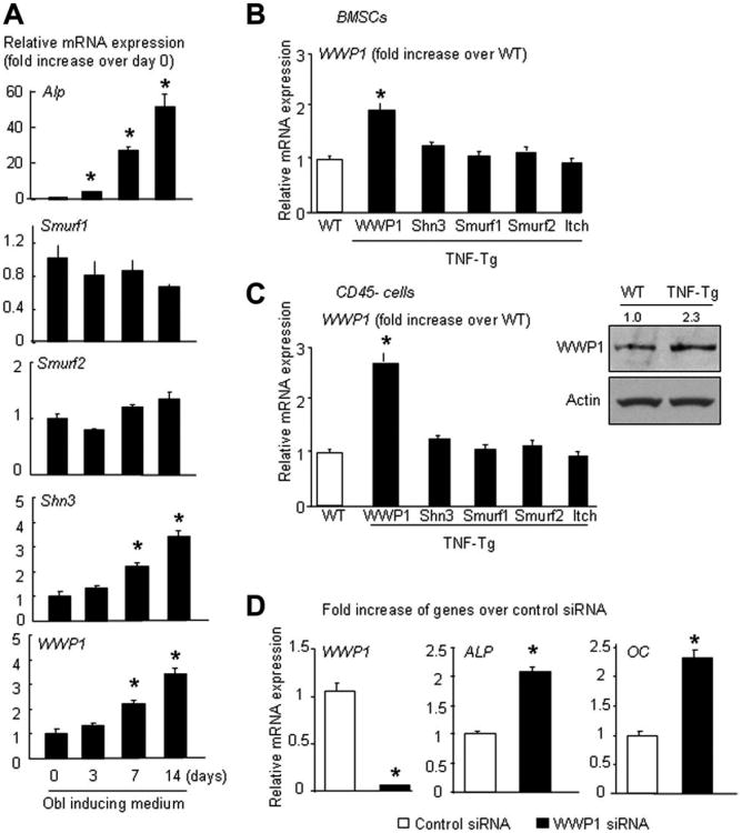
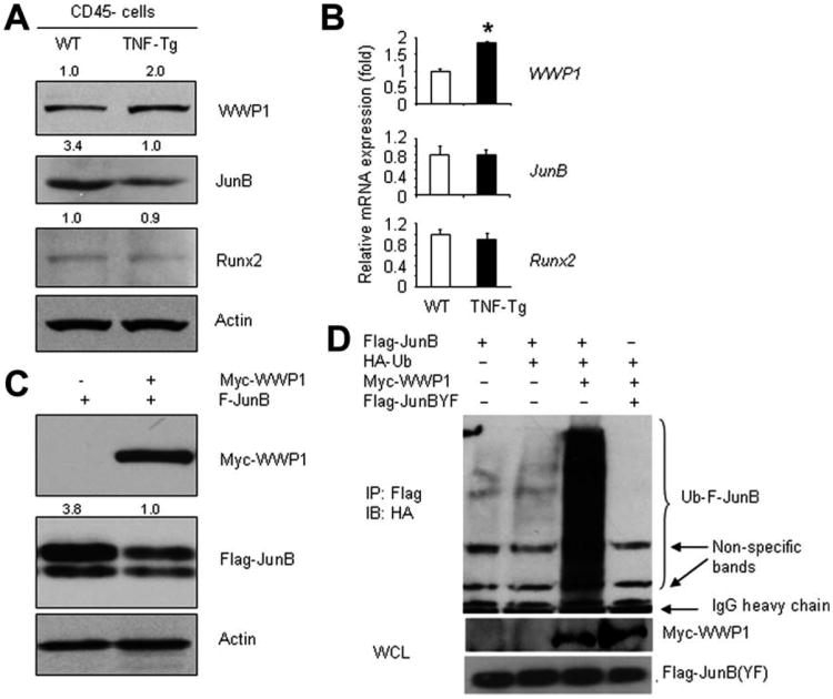
Patients with chronic inflammatory disorders, such as rheumatoid arthritis, often have osteoporosis due to a combination of Tumor necrosis factor-induced increased bone resorption and reduced bone formation. To test if TNF inhibits bone formation by affecting the commitment and differentiation of mesenchymal stem cells (MSCs) into osteoblasts, we examined the osteogenic potential of MSCs from TNF transgenic (TNF-Tg) mice, a model of chronic inflammatory arthritis. MSC-enriched cells were isolated from bone marrow stromal cells using negative selection with anti-CD45 antibody coated magnetic beads. The expression profile of MSC surface markers the osteogenic, chondrogenic, and adipogenic properties of CD45(-) cells were confirmed by FACS and cell differentiation assays. MSC-enriched CD45(-) cells from TNF-Tg mice formed significantly decreased numbers of fibroblast and ALP(+) colonies and had a decreased expression of osteoblast marker genes. As TNF may upregulate ubiquitin ligases, which negatively regulate osteoblast differentiation, we examined the expression levels of several ubiquitin ligases and found that Wwp1 expression was significantly increased in MSC-enriched CD45(-) cells of TNF-Tg mice. Wwp1 knockdown rescued impaired osteoblast differentiation of TNF-Tg CD45(-) cells. Wwp1 promotes ubiquitination and degradation of JunB, an AP-1 transcription factor that positively regulates osteoblast differentiation. Injection of TNF into wild-type mice resulted in decreased osteoblast differentiation of MSCs and increased JunB ubiquitination, which was completely blocked in Wwp1(-/-) mice. Thus, Wwp1 targets JunB for ubiquitination and degradation in MSCs after chronic exposure to TNF, and inhibition of Wwp1 in MSCs could be a new mechanism to limit inflammation-mediated osteoporosis by promoting their differentiation into osteoblasts.


Figures

Figure 1
Figure 1
TNF-Tg mice have decreased osteoblastic bone formation and mesenchymal colony formation. Serum bone formation and resorption markers were measured in 5- to 7-month-old TNF-Tg and wild-type (WT) mice by ELISA (enzyme-linked immunosorbent assay). Bars are the mean of SD of 18 mice per genotype (A). Representative images of fluorochrome-markers double labeled trabecular bone at distal metaphysic of femur in WT and TNF-Tg mice (B). Histomorphometric analysis. Bars are the mean of SD of eight mice per genotypes (C). Bone marrow stromal cells (BMSCs) from TNF-Tg mice and WT littermates (D, E) or from WT mice injected with TNF or PBS (F, G) were cultured in the osteoblast differentiation medium for 25–28 days in a colony formation assay. Cells were fixed and stained with eosin or for alkaline phosphatase (ALP) activity (D, F). Representative colony-forming unit (CFU)-ALP+ colonies are shown in (E, G). The numbers of CFU-F (eosin staining) and CFU-ALP+ colonies were counted and normalized to the WT or PBS-injected WT samples. Bars represent the mean ± SD of four dishes per genotype. (F): BMSCs from WT and TNF-Tg mice were cultured in the osteoblast differentiation medium and ALP and OC mRNA levels were examined by real time-polymerase chain reaction. Values shown are the mean ± SD of triplicates. The fold increase was calculated by dividing the values from different groups by the value of WT cells at day 0 as 1. *, p < .05 versus cells of WT or PBS-injected mice. The experiments were repeated three times with similar results. Abbreviations: ALP, alkaline phosphatase; BFR, bone formation rate; BS, bone surface; CFU, colony-forming unit; MAR, mineral apposition rate; MS, mineralizing surface; OC, osteocalcin; PBS, phosphate buffered saline; TNF, tumor necrosis factor; WT, wild-type.
Figure 2
Figure 2
Characterization of a mesenchymal stem cell (MSC)-enriched population derived from bone marrow stromal cells (BMSCs) of adult mice. Wild-type BMSCs were cultured in α-minimal essential medium, passaged twice, and subjected to flow cytometry after staining with FITC-anti-CD45, PE-anti CD105, biotin labeled hematopoietic lineage flow cocktail, and DAPI. (A): Scatter plots show that 99.2% of CD45cells are lineage negative and 52.8% of them are CD105+. (B): CD45+ hematopoietic lineage cells and CD45 MSC-enriched cells were purified using microbeads conjugated with an anti-CD45 antibody. Purified cells were cultured in osteoblast, adipocyte, or chondrocyte differentiation medium. Alkaline phosphatase (an osteoblast marker), Alizarin red (marker of matrix mineralization), oil red O (lipid), and Alcian Blue (chondrocyte) staining were performed. (C): CD45 cells were stained with PE-anti-CD105, allophycocyanin (APC)-anti-CD45, fluorescein isothiocyanate (FITC) -anti-CD11b, FITC-anti-CD31, PE-anti-CD29, APC-anti-CD44, PE-cy5-Sca-1, and FITC-anti-CD106 and then subjected to flow cytometry. The experiments were repeated once with similar results. Abbreviations: ALP, alkaline phosphatise; DAPI, 4′,6-diamidino-2-phenylindole.
Figure 3
Figure 3
Mesenchymal stem cell (MSC)-enriched CD45 cells derived from TNF-Tg mice have decreased osteoblast differentiation. (A): MSC-enriched CD45 cells isolated from BMSCs of wild-type (WT) mice were cultured in the osteoblast differentiation medium with or without TNF (7.5 ng/ml). Alkaline phosphatase (ALP) staining was performed after 4 days of culture. (B): CD45 MSC-enriched cells isolated from BMSCs of TNF-Tg mice and WT littermates were cultured in the osteoblast differentiation medium plus 20 ng/ml BMP-2 for 10 days. Cells were subjected to ALP staining and ALP activity measurement and (C) osteoblast marker gene expressions of CD45 cells were measured by real time-polymerase chain reaction. ALP activity was normalized to the total protein concentration for each sample. The relative ALP activity or gene expression levels was calculated by dividing the value from TNF-Tg cells by the values from WT cells which were normalized to 1. Values are the mean ± SD of four wells. *, p < .05 versus WT mice. The experiments were repeated three times with similar results. (D): TNF-Tg mice and WT littermates were injected with BrdU and harvested BMSCs were subjected to flow cytometry. The percentage of BrdU positive cells in the CD45 cells was calculated. The values are the mean + SD of six mice per genotype. Abbreviations: ALP, alkaline phosphatise; OC, osteocalcin; PBS, phosphate buffered saline; TNF, tumor necrosis factor; WT, wild-type.
Figure 4
Figure 4
Increased expression of the E3 ligase Wwp1 in bone marrow stromal cell (BMSCs) and MSC-enriched CD45 cells of TNF-Tg mice. (A): BMSCs harvested from wild-type (WT) mice were cultured in the osteoblast differentiation medium for the indicated time. The expression levels of E3 ligases were measured by real time-polymerase chain reaction (RT-PCR.) The fold increase was calculated by dividing the values of different groups by the value of day 0 which was normalized to 1. The values are the mean + SD of triplicates. (B, C): The expression levels of E3 ligases in BMSCs (B) and MSC-enriched CD45 cells (C) of TNF-Tg mice and WT littermates were determined by real time-polymerase chain reaction. The fold increase was calculated by dividing the value of the gene of interest by the value of the same gene from WT cells which was normalized to 1. The values are the mean + SD of triplicates. The Wwp1 protein level of CD45 cells was determined by Western blot analysis (inset). Fold change of Wwp1 protein was quantified using Image J and normalized to actin levels. (D): MSC-enriched CD45 cells from TNF-Tg mice were transfected with Wwp1 siRNA or control siRNA, and 2 days after transfection, cells were cultured in the osteoblast differentiation medium with BMP-2 (200 ng/ml) for 4 days. The expression levels of the gene of interest were examined by RT-PCR. The values are the mean + SD of three wells. The fold increase was calculated by dividing the values of Wwp1 siRNA-transfected cells by the value of control siRNA-transfected cells which was normalized to 1. *, p < .05 versus WT cells or control siRNA. The experiments were repeated three times with similar results. Abbreviations: ALP, alkaline phosphatase; BMSC, bone marrow stromal cell; OC, osteocalcin; siRNA, small interfering RNA; TNF, tumor necrosis factor; WT, wild-type.
Figure 5
Figure 5
Wwp1 induces JunB ubiquitination and degradation. (A): The expression levels of Wwp1, JunB, and Runx2 protein in mesenchymal stem cell (MSC)-enriched CD45 cells were examined by Western blot analysis. Fold changes were quantified using Image J and normalized to actin levels. (B): The expression levels of Wwp1, JunB and Runx2 mRNA in MSC-enriched CD45 cells were examined by real time-polymerase chain reaction (RT-PCR). The fold increase was calculated by dividing the values of TNF-Tg cells by the value of wild-type cells which was defined as 1. The values are the mean + SD of three wells. (C): 293T cells were transfected with Myc-Wwp1 and Flag-JunB expression plasmids for 48 hours. The expression of transfected Wwp1 and JunB was determined by anti-Myc and anti-Flag antibodies, respectively. Fold change of Flag-JunB was quantified using Image J and normalized to actin level. (D): 293T cells were transfected with Myc-Wwp1, hemagglutinin (HA)-ubiquitin, Flag-JunB, or JunB-PY mutant for 48 hours and treated with MG132 (10 μM) in the last 4 hours. Cell lysates were subjected to IP with anti-Flag antibody and blotted with anti-HA to detect ubiquitinated JunB protein. The experiments were repeated three times with similar results. Abbreviations: TNF, tumor necrosis factor; WT, wild-type.
Figure 6
Figure 6
TNF-induced mesenchymal stem cell (MSC) inhibition and JunB ubiquitination is abolished in Wwp1−/− mice. Wwp1−/− and wild-type (WT) mice were injected with either PBS or TNF as described in Figure 1. Bone marrow stromal cells (BMSCs) were subjected to colony-forming unit (CFU) assay, osteoblast marker gene expression, and JunB ubiquitination. (A): The numbers of CFU-F (eosin staining) colonies were counted and normalized to PBS-injected WT mice (which was set at 100%). Bars represent mean ± SD of four dishes per genotype. (B): ALP and OC mRNA levels of BMSCs from injected WT and Wwp1−/− mice were examined using quantitative RT-PCR. The values are mean ± SD of four dishes per genotype. (C): BMSCs derived from WT and Wwp1−/− mice injected with either PBS or TNF were treated with MG 132 (10 μM) for 4 hours and then lysed for ubiquitination assays to detect endogenous ubiquitinated JunB proteins. *, p < .05 versus PBS-injected WT. n = 4 per genotype. Abbreviations: ALP, alkaline phosphatase; CFU, colony-forming unit; OC, osteocalcin; PBS, phosphate buffered saline; TNF, tumor necrosis factor; WT, wild-type.

Similar articles

Cited by

References

    1. Canalis E. Effects of tumor necrosis factor on bone formation in vitro. Endocrinology. 1987;121:1596–1604. - PubMed
    1. Li YP, Stashenko P. Proinflammatory cytokines tumor necrosis factor-alpha and IL-6, but not IL-1, down-regulate the osteocalcin gene promoter. J Immunol. 1992;148:788–794. - PubMed
    1. Nanes MS. Tumor necrosis factor-alpha: Molecular and cellular mechanisms in skeletal pathology. Gene. 2003;321:1–15. - PubMed
    1. Gilbert L, He X, Farmer P, et al. Expression of the osteoblast differentiation factor RUNX2 (Cbfa1/AML3/Pebp2alpha A) is inhibited by tumor necrosis factor-alpha. J Biol Chem. 2002;277:2695–2701. - PubMed
    1. Diarra D, Stolina M, Polzer K, et al. Dickkopf-1 is a master regulator of joint remodeling. Nat Med. 2007;13:156–163. - PubMed

Publication types

MeSH terms