. 2011 Aug 23;124(8):876-85.
doi: 10.1161/CIRCULATIONAHA.110.005405. Epub 2011 Aug 1.

Genetic variation in titin in arrhythmogenic right ventricular cardiomyopathy-overlap syndromes

Affiliations

Genetic variation in titin in arrhythmogenic right ventricular cardiomyopathy-overlap syndromes

Matthew Taylor et al. Circulation. .

Abstract

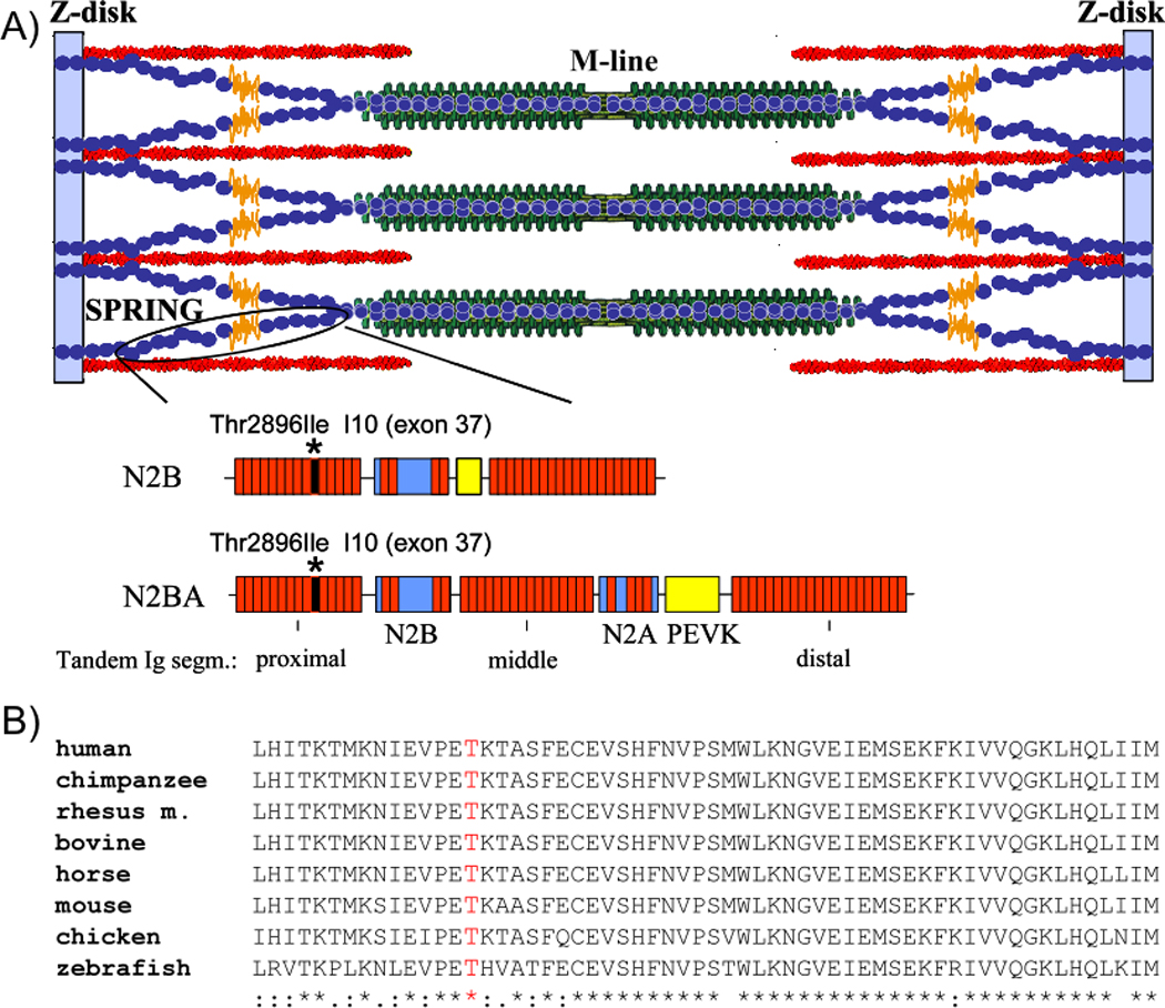
Background: Arrhythmogenic right ventricular cardiomyopathy (ARVC) is an inherited genetic myocardial disease characterized by fibrofatty replacement of the myocardium and a predisposition to cardiac arrhythmias and sudden death. We evaluated the cardiomyopathy gene titin (TTN) as a candidate ARVC gene because of its proximity to an ARVC locus at position 2q32 and the connection of the titin protein to the transitional junction at intercalated disks.

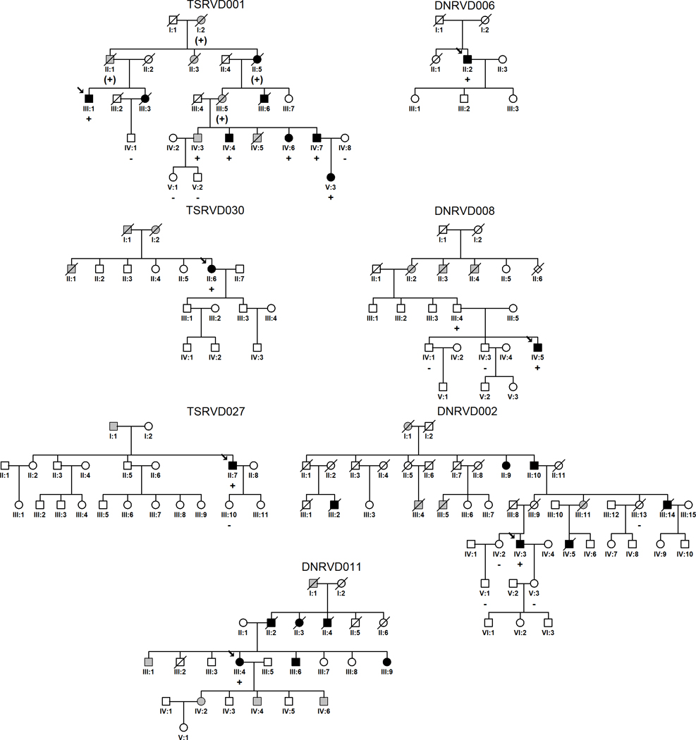
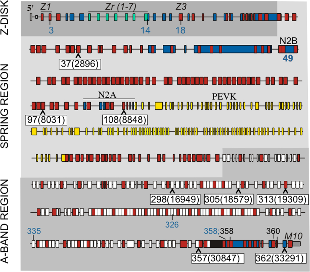
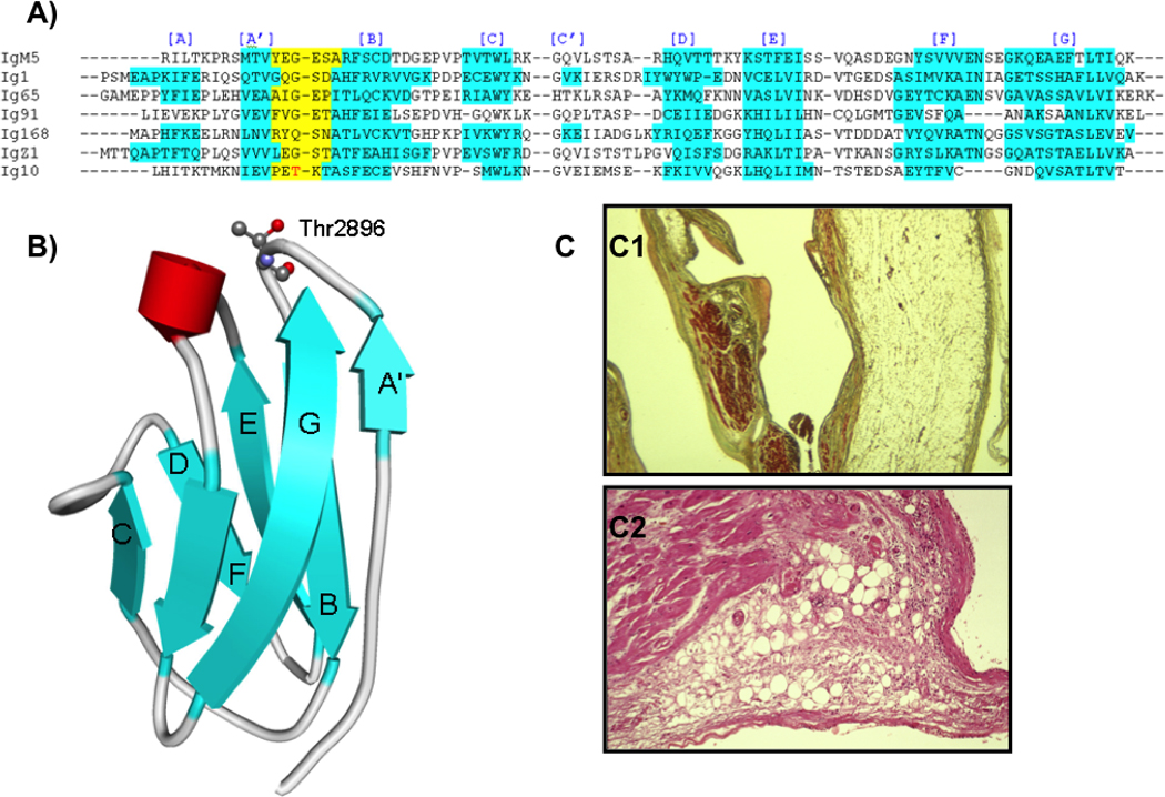
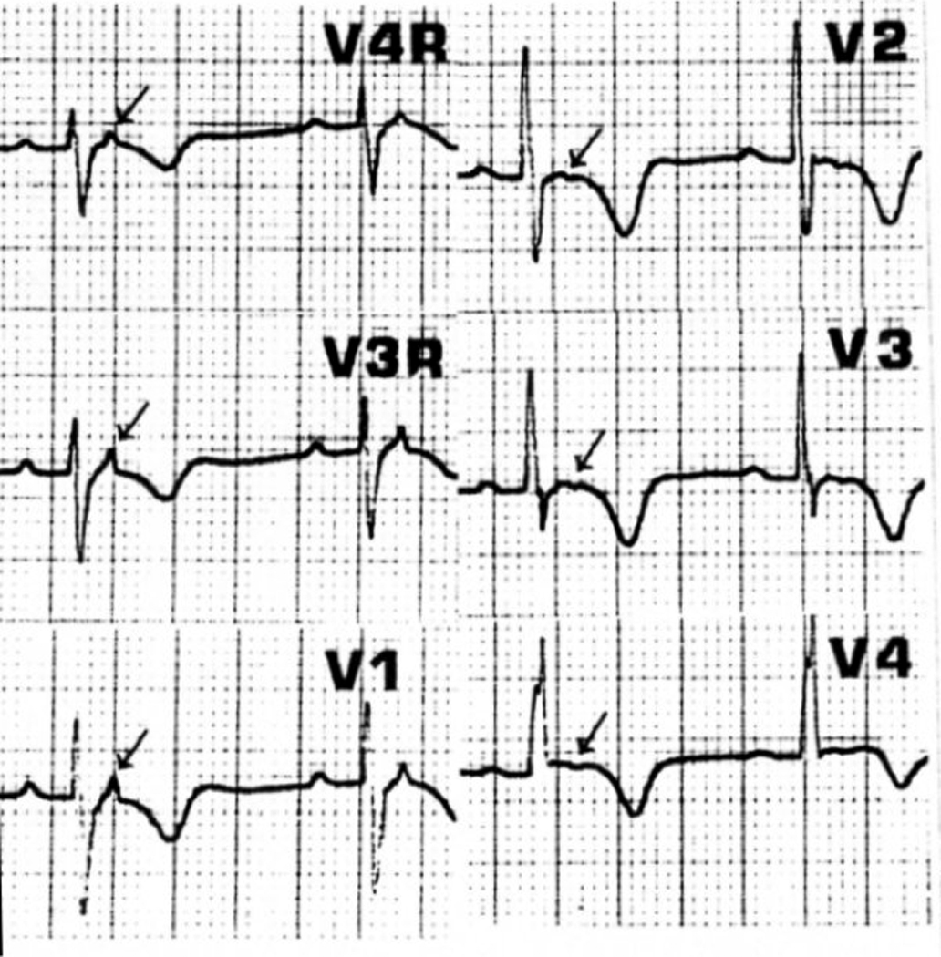
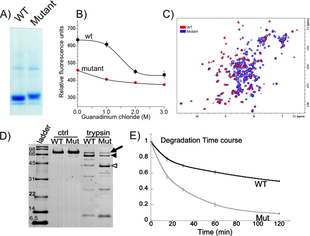
Methods and results: All 312 titin exons known to be expressed in human cardiac titin and the complete 3' untranslated region were sequenced in 38 ARVC families. Eight unique TTN variants were detected in 7 families, including a prominent Thr2896Ile mutation that showed complete segregation with the ARVC phenotype in 1 large family. The Thr2896IIe mutation maps within a highly conserved immunoglobulin-like fold (Ig10 domain) located in the spring region of titin. Native gel electrophoresis, nuclear magnetic resonance, intrinsic fluorescence, and proteolysis assays of wild-type and mutant Ig10 domains revealed that the Thr2896IIe exchange reduces the structural stability and increases the propensity for degradation of the Ig10 domain. The phenotype of TTN variant carriers was characterized by a history of sudden death (5 of 7 families), progressive myocardial dysfunction causing death or heart transplantation (8 of 14 cases), frequent conduction disease (11 of 14), and incomplete penetrance (86%).

Conclusions: Our data provide evidence that titin mutations can cause ARVC, a finding that further expands the origin of the disease beyond desmosomal proteins. Structural impairment of the titin spring is a likely cause of ARVC and constitutes a novel mechanism underlying myocardial remodeling and sudden cardiac death.


Figures

Figure 1
Figure 1
Pedigrees of ARVC families with rare TTN variants. Males and females are indicated by squares and circles respectively. Individuals meeting full ARVC diagnostic criteria are indicated with black shading; grey shading indicates a suggestive cardiac history and/or history of sudden unexplained death (see also Table 2). White squares/circles indicate unaffected individuals based on available family and medical history or, when TTN variant status is indicated, on the basis of full clinical evaluation by the investigators. TTN rare variant status for tested individuals is indicated by ‘+’ (present) and ‘-’ (absent) symbols; parentheses indicates inferred status. Probands are identified with an arrow.
Figure 2
Figure 2
Exon structure of human titin gene with location of rare TTN variants in ARVC families (black font on white background) indicated by exon location and amino acid change (in parentheses). Shown are also previously identified variants associated with other cardiac diseases. Variants in exons 3, 14 (2 different variants), 18, 49 (4 different variants), 326, 335, and 358 have all been associated with dilated cardiomyopathy (blue); additional variants in exons 358 and 360 have been associated with fetal cardiomyopathy (black). For details, original citations, and variants associated with skeletal muscle myopathies, see. (Red rectangle: immunoglobulin-like domain; white: fibronectin type 3 domain; blue: unique sequence; green: z-repeat domain; yellow: PEVK domain; black: titin kinase domain. (Figure based on Genbank accession AJ277892 and Bang et al.)
Figure 3
Figure 3
A) Multiple sequence alignments with known Ig structures, indicate that the mutated threonine in Ig10 is located in the short loop connecting the A' and B β-strands. The hydrogen bond network between the A' and G strands is important for Ig mechanical stability, and mutations in the A'B loop have been shown to disrupt this stabilizing network., B) Schematic representation of human Ig10 using the homology modeling server ModWeb with Thr2896, shown with a ball-and-stick model. C) Histological section of the right ventricular wall of patient III-3 from family TSRVD001 (Thr2896Ile). C1. The myocardium is substituted by fatty tissue and a layer of subendocardial fibrous tissue (Azan; original magnification x2.5). C2. Fibro-fatty infiltration with presence of inflammatory cells (Hematoxilin-eosin; original magnification x4.5.)
Figure 4
Figure 4
Electrocardiogram from individual IV-7 from family TSRVD001 (Thr2896Ile) with arrows indicating epsilon waves.
Figure 5
Figure 5
A) Schematic of titin in the sarcomere; sequences of spring regions of the two main cardiac isoforms (N2B and N2BA titin) are indicated schematically at bottom. In both isoforms the spring consists of tandemly arranged immunoglobulin-like domains (red), the N2B element and the PEVK. The N2BA isoform also contains the N2A element. The main ARVC mutation identified in this work (Thr2896Ile) is located in Ig10 (encoded by exon 37). B) Ig10, including T2896, is highly conserved in a wide range of species.
Figure 6
Figure 6
A) Reduced electrophoretic mobility of mutant Ig10 Thr2896Ile domain in native gel indicates a larger hydrodynamic radius, which is typical for unfolded proteins. B) Tryptophan fluorescence at various GuCl concentrations. Fluorescence in the absence of GuCl is much lower in mutant Ig10 than WT Ig10, but this difference is reduced as denaturant concentration increases. This suggests that under native conditions tryptophan is more exposed to hydrophilic conditions in mutant than WT Ig10. C) 15N HSQC NMR spectra of WT (red) and mutant Ig10 (blue). In the WT protein spectrum, we observe all expected peaks (one peak per backbone NH, Trp indole NH and two peaks per Gln/Asn NH2 moieties). The chemical shift dispersion is in accord with that expected for an IG domain fold protein. In contrast, the mutant exhibits at least two sets of NMR signals: one set with chemical shifts (and structure) similar to that of the WT domain and one or more additional sets with less chemical shift dispersion, which indicates the presence of additional unstructured molecules in the mutant domain sample. D) SDS-PAGE of recombinant WT and mutant Ig 7–13 fragments (I10 with Thr2896Ile mutation), in the absence of trypsin (ctrl) and after one hour of trypsin incubation (~25:1 Ig:trypsin by weight). The full-length peptides (arrow) have a MW of ~75 kDa. Trypsin treatment causes severe proteolysis and the appearance of a ~ 60 kDa fragment (filled arrowhead) which likely reflects the cleavage of one of the terminal Ig domains. In addition there is a prominent ~35–40 kDa degradation product (open arrowhead) that must result from a cleavage site somewhere in the middle of the Ig 7–13 protein; this fragment is much more prominent in the mutant sample. A protein MW ladder is shown for reference. E) Ratio of large Ig fragments (full-length protein + 60 kDa fragment) to all Ig fragments, by OD, as a function of trypsin incubation time. Degradation is more severe in the mutant protein. Error bars: ± SE; n = 4 for all time points except t = 120 min (n =3).

References

    1. Awad MM, Dalal D, Tichnell C, James C, Tucker A, Abraham T, Spevak PJ, Calkins H, Judge DP. Recessive arrhythmogenic right ventricular dysplasia due to novel cryptic splice mutation in PKP2. Hum Mutat. 2006;27:1157. - PMC - PubMed
    1. Basso C, Corrado D, Marcus FI, Nava A, Thiene G. Arrhythmogenic right ventricular cardiomyopathy. Lancet. 2009;373:1289–1300. - PubMed
    1. McKenna WJ, Thiene G, Nava A, Fontaliran F, Blomstrom-Lunqvist C, Fontaine G, Camerini F. Diagnosis of arrhythmogenic right ventricular dysplasia/cardiomyopathy. Br. heart J. 1994;71:215–218. - PMC - PubMed
    1. Nava A, Thiene G, Canciani B, Scognamiglio R, Daliento L, Buja G, Maertini B, Stritoni P, Fasoli G. Familial occurrence of right ventricular dysplasia. A study involving nine families. J Am Coll Cardiol. 1988;12:1222–1228. - PubMed
    1. den Haan AD, Tan BY, Zikusoka MN, Llado LI, Jain R, Daly A, Tichnell C, James C, Amat-Alarcon N, Abraham T, Russell SD, Bluemke DA, Calkins H, Dalal D, Judge DP. Comprehensive desmosome mutation analysis in north americans with arrhythmogenic right ventricular dysplasia/cardiomyopathy. Circ Cardiovasc Genet. 2009;2:428–435. - PMC - PubMed

Publication types

MeSH terms