. 2011 Sep 14;52(10):7195-204.
doi: 10.1167/iovs.11-7584.

The pattern of learned visual improvements in adult amblyopia

Affiliations

The pattern of learned visual improvements in adult amblyopia

Andrew T Astle et al. Invest Ophthalmol Vis Sci. .

Abstract

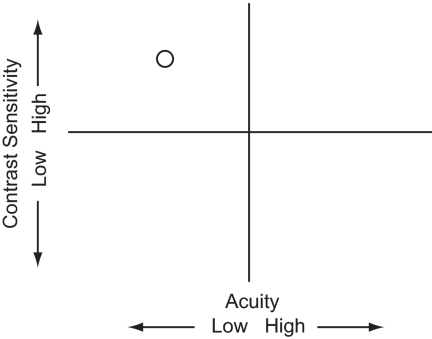
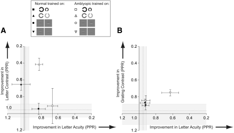
Purpose: Although amblyopia is diagnosed in terms of a monocular letter acuity loss, individuals typically present with deficits on a wide range of spatial tasks. Many of these deficits can be collapsed along two basic visual dimensions (visual acuity and contrast sensitivity) that together account for most of the variability in performance of the amblyopic visual system. In this study, this space was exploited, to target the main deficits and fully characterize the pattern of learned visual improvements in adult amblyopic subjects.

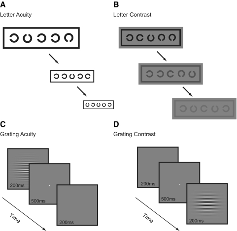
Methods: Twenty-six amblyopic subjects (mean age, 39 ±12 years) were trained on one of four tasks, categorized as either visual acuity (letter or grating acuity) or contrast sensitivity (letter or grating contrast) tasks. Performance was measured on all tasks before and after training, to quantify learning along each dimension and generalization to the other dimension. Performance in 35 visually normal subjects (mean, age 24 ± 5 years) was used to establish normal variation in visual performance along each dimension, against which the learned improvements in amblyopic subjects was compared.

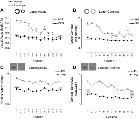
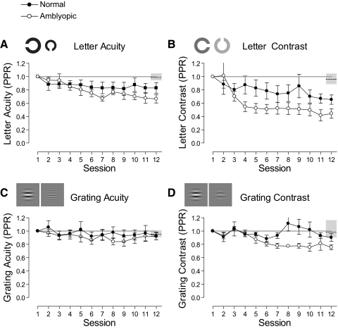
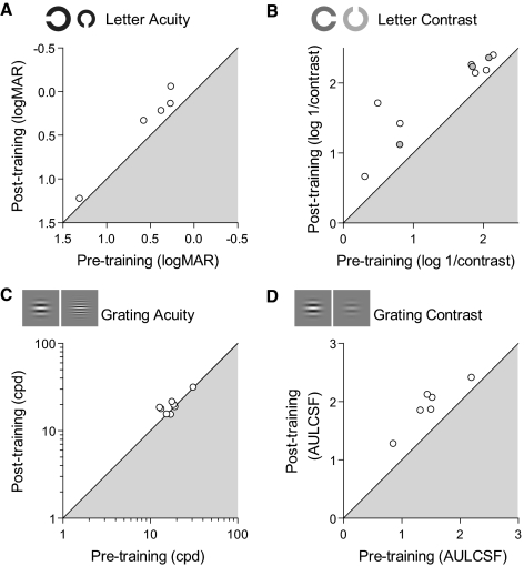
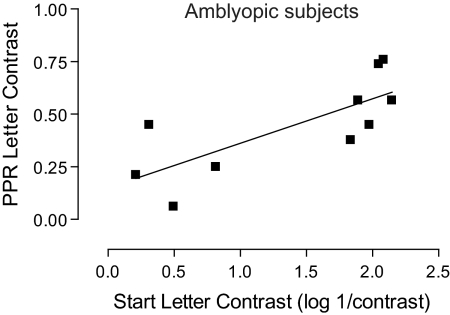
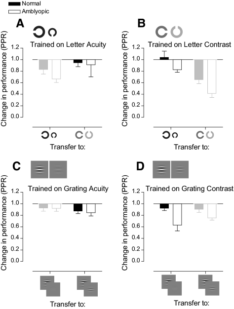
Results: Training on the contrast sensitivity tasks produced substantial within-task learning and generalization to measures of visual acuity. The learned improvements in performance after training on the letter acuity task were also substantial, but did not generalize to contrast sensitivity.

Conclusions: Mapping the pattern of learning onto the known deficit space for amblyopia enabled the identification of tasks and stimulus configurations that optimized learning, guiding further development of learning-based interventions in this clinical group.


Figures

Figure 1.
Figure 1.
Acuity and sensitivity deficit space. This space consists of two orthogonal dimensions: visual acuity, and contrast sensitivity. These two factors account for most of the variance in amblyopic visual performance, and individuals with different presumed etiologies occupy unique positions in this space. The amblyopic subject plotted here had poor visual acuity but relatively good contrast sensitivity.
Figure 2.
Figure 2.
The four computer-based psychophysical tasks used to assess performance along the acuity and sensitivity dimensions. There are two tests of visual acuity (letter acuity and grating acuity) and two of contrast sensitivity (letter contrast and grating contrast).
Figure 3.
Figure 3.
Task CIs. Mean test and retest measurements for each of the tasks. Error bars show the lower (2.5%) and upper (97.5% percentile) bounds of each CI.
Figure 4.
Figure 4.
Example learning curves for normal and amblyopic subjects for each of the four trained tasks. Thresholds are plotted as a function of training session. The ordinates have been oriented so that points plotted closer to the bottom of the graphs refer to sessions with better performance. Open and closed circles indicate subjects with amblyopic and normal data, respectively. Dashed horizontal lines: mean performance of normal subjects who did not undergo training on the task. Shaded regions denote the 95% CIs for the task. Error bars, SEM.
Figure 5.
Figure 5.
Normalized mean learning curves for each of the tasks. Mean normalized performance is shown for each of the tasks for subjects with normal (●) and amblyopic (○) vision. Learned improvements in performance are expressed relative to performance on the first day of training. Points lying below the solid horizontal line (PPR = 1) indicate an improvement beyond the level of performance on the first day of training. Dashed horizontal lines: mean test–retest ratio of normal subjects who did not undergo training on the task (gray shaded region shows 95% CI). Error bars, SEM.
Figure 6.
Figure 6.
Performance for individual amblyopic subjects as a function of performance before training. Subjects with results lying on the diagonal showed no benefits from training. Axes have been oriented such that points lying outside the shaded region denote subjects whose performance improved on the task after training. (B) Gray filled circles correspond to subjects previously trained on the grating task.
Figure 7.
Figure 7.
Improvement in letter contrast plotted against start letter contrast, for amblyopic subjects trained on the letter contrast task with linear regression curve fit. The two variables correlated strongly (r8 = 0.76; P < 0.05).
Figure 8.
Figure 8.
Bar charts showing improvements in performance on the trained task and transfer of learning to the untrained task. Each bar graph shows data for subjects who trained on one of the tasks. For example, (A) shows data for subjects who trained on the letter acuity task. It shows improvements on the trained task (letter acuity) and transfer of improvements in performance to the letter contrast task for these subjects. Results corresponding to within-task learning are displayed in lower contrast.
Figure 9.
Figure 9.
Improvements in performance mapped onto acuity–contrast space. Improvements are expressed as changes in performance along two dimensions: acuity (abscissa) and contrast (ordinate). (A) Data for those trained on letter tasks. (B) Grating task data. Horizontal dashed line: the mean letter contrast (A) or grating contrast (B) improvement for normal subjects who did not train (shaded regions straddling these lines show 95% CIs). Vertical dashed line: the letter (A) or grating (B) acuity improvement for normal subjects who did not train (straddling shaded regions shows 95% CIs). Axes are oriented such that points lying away from the origin and outside the CIs denote an improvement. Error bars represent the SEM.

References

    1. Ciuffreda K, Levi DM, Selenow A. Amblyopia: Basic and Clinical Aspects. Stoneham, MA: Butterworth-Heinemann; 1991
    1. von Noorden GK. Binocular Vision and Ocular Motility: Theory and Management of Strabismus. 5th ed St. Louis: Mosby; 1996
    1. Attebo K, Mitchell P, Smith W. Visual acuity and the causes of visual loss in Australia. The Blue Mountains Eye Study. Ophthalmology. 1996;103:357–364 - PubMed
    1. Stewart CE, Fielder AR, Stephens DA, Moseley MJ. Design of the Monitored Occlusion Treatment of Amblyopia Study (MOTAS). Br J Ophthalmol. 2002;86:915–919 - PMC - PubMed
    1. Wiesel TN, Hubel DH. Single-cell responses in striate cortex of kittens deprived of vision in one eye. J Neurophysiol. 1963;26:1003–1017 - PubMed

Publication types