. 2011 Sep 1;52(9):7001-6.
doi: 10.1167/iovs.11-7619.

Diffusion tensor imaging detects retinal ganglion cell axon damage in the mouse model of optic nerve crush

Affiliations

Diffusion tensor imaging detects retinal ganglion cell axon damage in the mouse model of optic nerve crush

Xu Zhang et al. Invest Ophthalmol Vis Sci. .

Abstract

Purpose: Diffusion tensor imaging (DTI) measures the random motion of water molecules reflecting central nervous system tissue integrity and pathology. Glaucoma damages retinal ganglion cells (RGCs) and their axons. The authors hypothesized that DTI-derived axonal and myelin injury biomarkers may be used to detect early axonal damage and may be correlated with RGC loss in the mouse model of optic nerve crush (ONC).

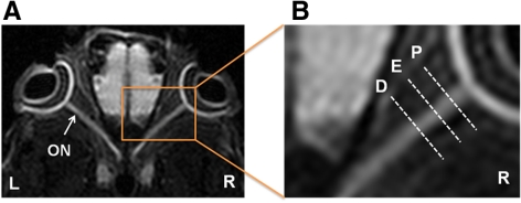
Methods: The progression of RGC axon degeneration was quantitatively assessed with DTI in vivo, corroborated with axon/myelin immunohistochemical staining and retrograde fluorogold labeling in mice after ONC.

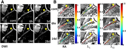
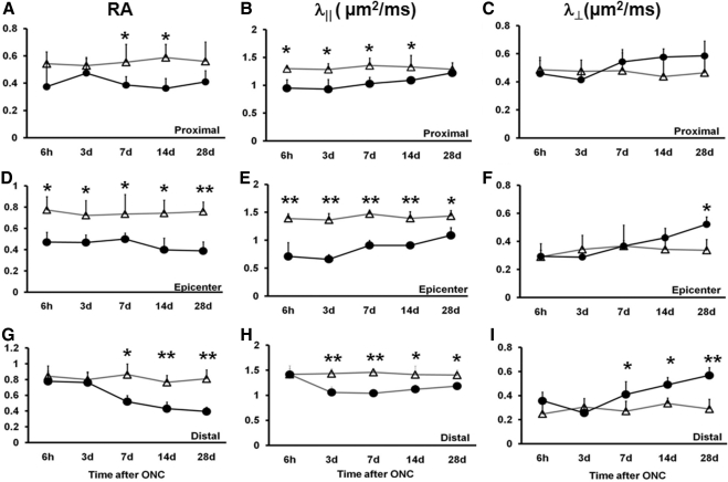
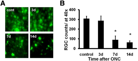
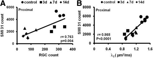
Results: Decreased axial diffusivity (λ(‖)) and relative anisotropy (RA) of damaged axons were observed from 6 hours to 14 days, reflecting axonal injury. DTI detected axonal injury at 6 hours after ONC when SMI-31 did not detect axonal abnormality. Both decreased λ(‖), and SMI-31 identified axon damage at 3 days after ONC. Decreased λ(‖) correlated with reduced SMI-31-positive axon counts from 3 days after ONC. In contrast, the increased λ(⊥) was seen only in the distal segment of optic nerve whereas decreased myelin basic protein-positive axon counts were seen in all segments 3 days after ONC. The number of retrograde-labeled RGCs did not decline significantly until 7 days after ONC. There was a significant correlation between RGC loss and optic nerve axon damage.

Conclusions: The authors demonstrated that in vivo DTI detected axonal injury earlier than SMI-31. Results suggest that in vivo DTI of optic nerve injury may be used as a noninvasive tool for assessing the pathogenesis of RGC axonal injury.


Figures

Figure 1.
Figure 1.
Defining ROI in a control DWI map. Both eyeballs (L, left eye; R, right eye) and optic nerves (ON) are clearly seen in DWI. The ROI is defined as follows: proximal (P), 300 to 600 μm posterior to the globe; epicenter (E), approximately 900 to 1100 μm from the globe; distal (D), 1200 to 1600 μm posterior to the globe.
Figure 2.
Figure 2.
Serial in vivo diffusion MRI of the crushed optic nerve from a mouse (A, DWI; B, DTI maps). Yellow arrowheads: epicenter. The optic nerve is hyperintense in DWI (A). Increased intensity is seen at the epicenter 6 hours after ONC. Optic nerve ROI was defined based on the DWI (Fig. 1). Significantly decreased RA resulting from the axonal injury extended the entire ON at 14 days (B). Progressive myelin damage reflected as the increased λ is also clearly demonstrated (B). Decreased λ indicative of axonal injury after ONC clearly identified the epicenter (B). Image scales are as follow: RA, 0.0–1.0 (no unit); λ, 0.0–0.5 (μm2/ms); λ, 0.0–1.5 (μm2/ms).
Figure 3.
Figure 3.
The time course of DTI parameters measured from longitudinal analysis of crush-injured optic nerves (circles) and uninjured optic nerves (triangles). RA, λ, and λ of proximal, epicenter, and distal ROI are displayed as mean ± SD (n = 5). At the proximal site, the RA value decreased from day 7 (A), λ values significantly decreased from 6 hours (B), and λ increased around day 14 (C). At the epicenter, significant decreases in RA and λ were seen at all time points (D, E). Significantly increased λ was seen only at 28 days (F). At the distal site, a changed RA value was observed from day 7 (G), significantly decreased λ started at day 3 (H), and λ was significantly increased at day 7 (I). Statistical differences indicated in relation to control group: *P < 0.05, **P < 0.01, two-way ANOVA.
Figure 4.
Figure 4.
Immunohistochemistry of SMI-31 and MBP of the control and the crush-injured optic nerves. (A) SMI-31 of proximal optic nerve cross-sections from control (cont) and ONC injury mice at 3, 7, and 14 days. (B) Quantitative SMI-31–positive axon counts of the optic nerve at proximal (P), crushed epicenter (E), and distal (D) sites. (C) MBP of the proximal optic nerve cross-sections from the control (cont) and ONC-injured mice at 3, 7, and 14 days after injury. (D) Quantitative MBP-positive axon counts at the three selected sites. The timing of myelin injury reflected as loss of MBP-positive axon counts is similar to that of SMI-31. *P < 0.05, **P < 0.01. Magnification, 60×.
Figure 5.
Figure 5.
RGCs were retrogradely labeled with FG a week before ONC. Flat-mount retinas, at 40× magnification, were examined at 3, 7, and 14 days after ONC (A). Quantitative estimation of the remaining RGCs was performed after ONC using fluorescence microscopy (mean ± SEM; n = 3 for each experimental group; B). Crush injury induced a significant loss of RGCs at both 7 and 14 days after ONC. *P < 0.05.
Figure 6.
Figure 6.
Correlations of the SMI-31 counts with RGC loss (A) and axial diffusivity (B) after ONC. FG-labeled RGC counts correlated with the numbers of SMI-31–stained axons. Axial diffusivity also correlated with SMI-31 axon counts at the proximal site.

Similar articles

Cited by

References

    1. Quigley HA, Broman AT. The number of people with glaucoma worldwide in 2010 and 2020. Br J Ophthalmol. March 2006. 2006;90(3):262–267 - PMC - PubMed
    1. Quigley HA, Dunkelberger GR, Green WR. Retinal ganglion cell atrophy correlated with automated perimetry in human eyes with glaucoma. Am J Ophthalmol. 1989;107(5):453–464 - PubMed
    1. Buckingham BP, Inman DM, Lambert W, et al. Progressive ganglion cell degeneration precedes neuronal loss in a mouse model of glaucoma. J Neurosci. 2008;28(11):2735–2744 - PMC - PubMed
    1. Crish SD, Sappington RM, Inman DM, Horner PJ, Calkins DJ. Distal axonopathy with structural persistence in glaucomatous neurodegeneration. Proc Natl Acad Sci U S A. 2010;107(11):5196–5201 - PMC - PubMed
    1. Schlamp CL, Li Y, Dietz JA, Janssen KT, Nickells RW. Progressive ganglion cell loss and optic nerve degeneration in DBA/2J mice is variable and asymmetric. BMC Neurosci. 2006;7:66. - PMC - PubMed

Publication types