. 2011 Jul;9(7):e1001107.
doi: 10.1371/journal.pbio.1001107. Epub 2011 Jul 19.

Synaptic transmission from horizontal cells to cones is impaired by loss of connexin hemichannels

Affiliations

Synaptic transmission from horizontal cells to cones is impaired by loss of connexin hemichannels

Lauw J Klaassen et al. PLoS Biol. 2011 Jul.

Abstract

In the vertebrate retina, horizontal cells generate the inhibitory surround of bipolar cells, an essential step in contrast enhancement. For the last decades, the mechanism involved in this inhibitory synaptic pathway has been a major controversy in retinal research. One hypothesis suggests that connexin hemichannels mediate this negative feedback signal; another suggests that feedback is mediated by protons. Mutant zebrafish were generated that lack connexin 55.5 hemichannels in horizontal cells. Whole cell voltage clamp recordings were made from isolated horizontal cells and cones in flat mount retinas. Light-induced feedback from horizontal cells to cones was reduced in mutants. A reduction of feedback was also found when horizontal cells were pharmacologically hyperpolarized but was absent when they were pharmacologically depolarized. Hemichannel currents in isolated horizontal cells showed a similar behavior. The hyperpolarization-induced hemichannel current was strongly reduced in the mutants while the depolarization-induced hemichannel current was not. Intracellular recordings were made from horizontal cells. Consistent with impaired feedback in the mutant, spectral opponent responses in horizontal cells were diminished in these animals. A behavioral assay revealed a lower contrast-sensitivity, illustrating the role of the horizontal cell to cone feedback pathway in contrast enhancement. Model simulations showed that the observed modifications of feedback can be accounted for by an ephaptic mechanism. A model for feedback, in which the number of connexin hemichannels is reduced to about 40%, fully predicts the specific asymmetric modification of feedback. To our knowledge, this is the first successful genetic interference in the feedback pathway from horizontal cells to cones. It provides direct evidence for an unconventional role of connexin hemichannels in the inhibitory synapse between horizontal cells and cones. This is an important step in resolving a long-standing debate about the unusual form of (ephaptic) synaptic transmission between horizontal cells and cones in the vertebrate retina.


Conflict of interest statement

The authors have declared that no competing interests exist.

Figures

Figure 1
Figure 1. Localization of cellular markers in retinas from wild-type and mutant zebrafish.
The organization of the outer retina was evaluated by the distribution of a number of cellular markers for double cones (A, FRet43) in red, horizontal cells (B, GluR2) in green, OFF-bipolar cells (C, GluR4) in red, and interplexiform cells (E, TH) in red. The blue stain is the nuclear marker DAPI. No difference in the distribution of these markers could be observed. In both preparations, GluR2 forms horseshoe-like structures in the OPL (B, D). Previous studies have shown that these structures are horizontal cell dendrites invaginating the cone terminal ,. GluR4 labeling is present as small puncta at the level of the OPL (C, D). These puncta were identified as the tips of the dendrites of OFF bipolar cells ,. (E) TH labels interplexiform cells and the synapses these cells make onto horizontal cells. The immunoreactivity pattern is similar in wild-type and mutant retinas. These results indicate that all major neuron types in the OPL have developed normally. (A to E): Scale bar = 10 µm. (F) Ultrastructure of the cone synaptic terminal in wild-type (left) and mutant (right) retinas. No differences were found in the ultrastructural organization of the cone synaptic terminal. In both animal models, the characteristic shape of the synaptic triad was easily identified. R, synaptic ribbon; Sp, spinules; HC, horizontal cell; * bipolar cell. Scale bar = 0.5 µm.
Figure 2
Figure 2. Cx52.9 co-localizes with GluR2, Cx55.5, and Cx52.6 in zebrafish retinas.
The top panel shows co-localization of the Cx52.9 (green) antibody and GluR2-IR (red). GluR2 labels the tips of horizontal cell dendrites. This co-localization shows that Cx52.9 is present at the horizontal cell dendrites. Middle panel: Double labeling of Cx52.9 (green) and Cx55.5 (red). Red, green, and yellow plaques can be found, indicating that in some (parts) of the gap-junctions Cx55.5 co-localizes with Cx52.9. The bottom panel shows that there is co-localization of Cx52.6 (red) and Cx52.9 (green). Also for this combination, areas of co-expression of the two connexins can be found. This means that the three connexins co-localize in gap-junctions between horizontal cells. Scale bar = 10 µm (top panel). Scale bar = 5 µm (middle and bottom panel).
Figure 3
Figure 3. Localization and expression of connexins expressed by horizontal cells in wild-type and mutant zebrafish.
(A) Immunocytochemical staining with antibodies against Cx55.5 (i), Cx52.6 (ii), and Cx52.9 (iii). Cx55.5-IR and Cx52.6-IR are absent in the mutant, whereas Cx52.9-IR remains present or even becomes stronger. Scale bar = 10 µm. (B) Double labeling of Cx52.9 (green) and FRet43 (red), a label for double cones. The Cx52.9-IR and FRet43-IR are closely associated, indicating that Cx52.9-IR is present in the dendrites of horizontal cells invaginating the cone synaptic terminal in both wild-type and mutant. Scale bar = 10 µm. (C) Tannic acid staining of gap-junctions in wild-type and mutant retinas. Pairs of arrow heads indicate the extent of the gap-junction. Often the gap-junctions in the mutant seem to be “split” as is illustrated in this figure. On average, gap-junctions are smaller in the mutant retinas. Scale bar = 0.5 µm.
Figure 4
Figure 4. qPCR and western blot data confirm absence of Cx55.5 and Cx52.6.
(A) Immunoreactivity patterns of Cx55.5, Cx52.6, and Cx52.9 antibodies in membrane samples of wild-type and C54X mutant zebrafish retinas. On Western blots derived from 8–10% SDS-PA gradient gels on which membrane samples (40 µg of each) of wild-type and mutant zebrafish retinas were separated, the Cx55.5 and Cx52.6 detected a double band with a molecular weight in the expected range (black asterisk) in the wild-type, but not the mutant retina (black asterisk). In contrast, the expression of Cx52.9 protein appeared to be unaffected in the mutant (white asterisk) and a protein with the appropriate molecular weight of Cx52.9 was detected in both membrane samples, wild-type and mutant. (B) qPCR experiments revealed a significant down regulation of Cx55.5 and Cx52.6 mRNA in mutant fish (p<0.001). Steady state Cx52.9 mRNA levels were not affected.
Figure 5
Figure 5. Horizontal cell properties in wild-type and mutant zebrafish.
(A) Ihemi in wild-type (black) and mutants (red) differ significantly. Whole cell currents of a dissociated horizontal cell when stepped to −60 mV and +60 mV, respectively. In the mutant, the inward Ihemi at −60 mV is reduced, whereas the outward current at +60 mV is enhanced. (B) IV relation of Ihemi in wild-type and mutants. Ihemi is minimally affected around VHC in the dark (−30 mV, arrow), is reduced at negative potentials, and is increased at positive potentials. (C) Responses to 500 ms flashes of 200 µm spot and full-field stimuli. The spot/full-field ratio did not differ between wild-type and mutant. (D) Distribution of horizontal cell classes in wild-type and in mutant fish did not differ significantly. (E) Three examples of spectrally coded horizontal cell responses in wild-type and mutants. The retina was stimulated with 500 ms full-field flashes of 624, 525, and 465 nm light.
Figure 6
Figure 6. Feedback measured in cones is reduced in the mutant zebrafish.
(A) Light-induced feedback responses in wild-type (left) and mutant (right) zebrafish cones. Horizontal cells were hyperpolarized by full-field 1 s flashes of light while the recorded cone was saturated with a 20 µm spot of bright light. (B) IV relation of a cone ICa in control (green), 50 µM DNQX (red), and 30 µM KA (black) in wild-type (Bi, left) and mutant (Bii, left). Averaged DNQX- (red) and KA- (black) induced currents in wild-type (Bi, right) and mutants (Bii, right). The DNQX-induced current is significantly reduced in the mutant compared to the current in wild-type.
Figure 7
Figure 7. Model for ephaptic feedback.
(A) Schematic drawing of the cone synaptic terminal. HC, horizontal cell; BC, bipolar cell; ICa, cone Ca2+-current; Vext, potential in the synaptic cleft. (B) The feedback model formulated by Fahrenfort et al. was used to evaluate the effect of changes in voltage dependence of horizontal cell hemichannels. Here we will give a short description of the main features, for a full description see Fahrenfort et al. . The model consists of a simple resistive network. The model was used to evaluate the relation between the horizontal cell membrane potential (VHC) and the change in extracellular potential (Vext) with different numbers of hemichannels in the horizontal cell membrane. Vext is the potential in the synaptic cleft, VHC is the horizontal cell membrane potential, ghemi is the hemichannel conductance, gGlu, tip is the glutamate conductance at the tips of the horizontal cell dendrites, gGlu,neuropil is the glutamate conductance of the horizontal cell dendrites in the neuropil, gK, is the potassium conductance of horizontal cells and EK is the equilibrium potential for potassium. [Glu] is the Glutamate-concentration in the synaptic cleft. ICa is the cone Ca2+-current; gext is the conductance of the extracellular space in the synaptic complex.
Figure 8
Figure 8. Model simulations of feedback induced shift of Ca-current in wild-type and mutant zebrafish.
Left: The simulated IV relations of formula image 
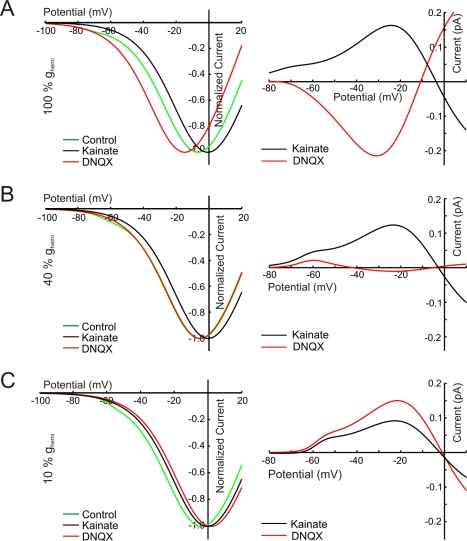
in control condition, DNQX, and KA, produced by the ephaptic feedback model . Right: the DNQX- and KA-induced feedback currents. The value of formula image was varied from 100% (A) to 10% (C) of the wild-type value . With reducing formula image, the DNQX-induced feedback currents decrease and finally reverse. The KA-induced feedback currents only decrease with decreasing formula image.
Figure 9
Figure 9. Optokinetic response of mutant zebrafish is reduced.
(A) Eye movements of a wild-type zebrafish larva (black, right eye; red, left eye). Timing of the stimulus is indicated in the bottom trace. (B) Optokinetic gain as function of contrast for 13 wild-type (black) and 13 mutant (red) zebrafish. Over the whole contrast range the wild-type performed significantly better than the mutant. (C) In the mutant, reduction in optokinetic gain is stronger for high temporal frequencies (1.0 cycle per second) than for low temporal frequencies (0.25 cycle per second) (p = 0.017). (D) In wild-type the temporal frequency transfer function of cones to bipolar cells is a band-pass filter. The low frequency cutoff is due to negative feedback from horizontal cells. When removing this pathway and inducing an overall gain reduction, the transfer function changes into a low-pass filter. At low frequencies this transformation leads to a smaller loss in gain than for high temporal frequencies.

Similar articles

Cited by

References

    1. Verweij J, Kamermans M, Spekreijse H. Horizontal cells feed back to cones by shifting the cone calcium-current activation range. Vision Res. 1996;36:3943–3953. - PubMed
    1. Thoreson W. B, Babai N, Bartoletti T. M. Feedback from horizontal cells to rod photoreceptors in vertebrate retina. J Neurosci. 2008;28:5691–5695. - PMC - PubMed
    1. Kamermans M, Fahrenfort I, Schultz K, Janssen-Bienhold U, Sjoerdsma T, Weiler R. Hemichannel-mediated inhibition in the outer retina. Science. 2001;292:1178–1180. - PubMed
    1. Hirasawa H, Kaneko A. pH changes in the invaginating synaptic cleft mediate feedback from horizontal cells to cone photoreceptors by modulating Ca2+ channels. J Gen Physiol. 2003;122:657–671. - PMC - PubMed
    1. Davenport C. M, Detwiler P. B, Dacey D. M. Effects of pH buffering on horizontal and ganglion cell light responses in primate retina: evidence for the proton hypothesis of surround formation. J Neurosci. 2008;28:456–464. - PMC - PubMed

Publication types