. 2011 Jul;7(7):e1002186.
doi: 10.1371/journal.pgen.1002186. Epub 2011 Jul 21.

Drosophila araucan and caupolican integrate intrinsic and signalling inputs for the acquisition by muscle progenitors of the lateral transverse fate

Affiliations

Drosophila araucan and caupolican integrate intrinsic and signalling inputs for the acquisition by muscle progenitors of the lateral transverse fate

Marta Carrasco-Rando et al. PLoS Genet. 2011 Jul.

Abstract

A central issue of myogenesis is the acquisition of identity by individual muscles. In Drosophila, at the time muscle progenitors are singled out, they already express unique combinations of muscle identity genes. This muscle code results from the integration of positional and temporal signalling inputs. Here we identify, by means of loss-of-function and ectopic expression approaches, the Iroquois Complex homeobox genes araucan and caupolican as novel muscle identity genes that confer lateral transverse muscle identity. The acquisition of this fate requires that Araucan/Caupolican repress other muscle identity genes such as slouch and vestigial. In addition, we show that Caupolican-dependent slouch expression depends on the activation state of the Ras/Mitogen Activated Protein Kinase cascade. This provides a comprehensive insight into the way Iroquois genes integrate in muscle progenitors, signalling inputs that modulate gene expression and protein activity.


Conflict of interest statement

The authors have declared that no competing interests exist.

Figures

Figure 1
Figure 1. Pattern of expression of ara and caup during myogenesis.
Wild type embryos of the indicated developmental stages were hybridized with caup (A, A′, B, B′, C, E, E′) or ara (F) riboprobes or sectioned after anti-Caup antibody staining (A″, A′″, B″, B′″, C′, D, D′, E″). (A-A′″) caup is expressed in the visceral mesoderm at early stage 11 (arrowheads, A and A′ show the same embryo with different focus as shown in the inset). (B-B′″) At mid stage 11 caup is expressed in the visceral mesoderm (arrowheads) and in the lateral ectoderm (arrows). Asterisks in A and B point to the primordium of the proventriculus. A′″, B′″ close-ups of the images shown in A″ and B″, respectively. (C-C′) Early stage 12/late stage 11 embryos. (C) caup is expressed in the lateral ectoderm (arrowhead) and in groups of mesodermal cells (arrow). (C′) Cross-section showing caup expression in ectodermal cells (Ec), visceral mesoderm (Vms) and promuscular clusters (Cl). (D, D′) At stage 12 caup is expressed in individual muscle progenitors (P in D) and slightly later in both founders (Fs) derived from the division of progenitors (D′). (E-E″) At stage 13 Caup is detected in a lateral stripe of ectodermal cells (arrowheads in E, E′, Ec in E″) and in muscle precursors (arrows in E, E′, M in E″). (F) Stage 15 embryo showing expression of ara in the ectoderm and in mature muscles. (G) Stage 15 embryos doubled stained with anti-Caup (green) and antibodies against Con, Slou or Ladybird (red). caup is co-expressed with Con in LT1–4 muscles, with slou in DT1 and with lb in SBM. The drawing scheme summarises the wild type patterns of expression of caup (green), slou (red), lb (yellow) and Con (black contour line) in relation to the wild type complement of abdominal muscles. For muscle nomenclature see .
Figure 2
Figure 2. Onset of Caup expression in muscles in relation to other muscle identity genes.
All images show a detail of an embryonic wild-type abdominal hemisegment stained with antibodies against Caup (green) and different muscle identity proteins. Images show a ventral view of the embryo, with the exception of B and C that correspond to lateral views. (A–C) Stage 11 embryos. (A) caup and Kr (red) are co-expressed in a lateral transverse promuscular cluster (CLTS). (B–C) caup is co-expressed with Kr (blue) in progenitors of LT muscles (PLT1/LT2 and PLT3/LT4, B) and with slou/S59 (red) in DT1/DO3 progenitor (PDT1/DO3, C). (D) Late stage 12 embryo co-expressing caup and lb in the SBM founder (FSBM). (E) Stage 12 embryo showing co-expression of caup with slou/S59 (red) in DT1 founder (FDT1) and with Kr (blue) in LTs founders (FLT1–4). The position of LL1, LL1sib and VA1–3 founders (FLL1, FLL1sib, FVA1–3) and the ventral adult muscle precursor are also indicated. (F) Schematic representation of ara/caup expression in the LTs, DT1 and SBM lineages (SBM lineage as revised in [17]). LaPs, lateral adult muscle precursors; PC, promuscular cluster; P, muscle progenitor; Fs, founder myoblasts.
Figure 3
Figure 3. Muscle phenotypes of Iro-C mutant embryos.
(A–D) Latero-ventral region of stage 16 wild-type (A), Df(3L)iroDFM3 (B), Df(3L)iroEGP6 (C) and stage 15 Df(3L)iroDFM3 mef-2GAL4::UAS-ara (D) embryos stained with anti- Tropomyosin antibody (green). The position of ventral wild-type LT muscle tips and LT attachment sites are marked with arrowheads and brackets, respectively. Note the absence of muscles with LT morphology and insertions at LT attachment sites, and the presence of morphologically normal DT1 and SBM muscles (arrows) in the mutant backgrounds (B, C). Asterisks indicate morphological abnormal latero-ventral muscles in these embryos. This phenotype is rescued by mesodermal ara expression with the pan-mesodermal driver mef2-GAL4 (D). (E) Quantification of phenotypes produced by the loss of ara/caup in LT muscles. * Refers to changes in shape, orientation or attachment sites; n, numbers of hemisegments analysed (stages 14–16); -, not determined.
Figure 4
Figure 4. Changes of fate in LT founders of Iro-C mutant embryos.
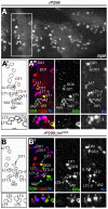
(A-B′) Late stage 12 control (A-A″) and rP298;;Df(3L)iroEGP6 sibling embryos (B, B′) stained with anti-ßgal (red), anti-Slou/S59 (green) and anti-Kr (blue) antibodies. ßgal staining is used as a marker for founders (rP298 line) and the white rectangle in A marks the individual segment shown in A′, A″. (A′, B) Drawings indicating the relative position of the founders visualised in the corresponding (A″, B′) confocal images. The founders expressing Kr or slou/S59 are labelled by their muscle's acronyms. Note that although founder segregation is unaffected in Df(3L)iroEGP6 embryos, the specification of LT founders is altered (B, B′). Thus, two of the LT founders (LT3–4* in B, B′), marked by expression of Kr, also express slou/S59, a property exclusive of the VA1–2 founders (see insets below for details of LT founders, the asterisks mark VT1 founder, that expresses slou/S59 but not Kr. Note that Kr is disappearing from LT1 and 3). All panels show Z projections of several consecutive confocal sections with the exception of A″ that corresponds to a combination of two Z projections, one lateral, as the one shown in B′, and other rotated ventrally to show VAs founders. For muscle nomenclature other than ventral adult precursor (VaP) see .
Figure 5
Figure 5. Muscle fate transformations in Df(3L)iroDFM3 embryos.
(A) Summary of identity codes for promuscular clusters (Cl), progenitors (P) and muscles missing or duplicated in ara/caup mutants, indicated by a colour code. (B) Schematic drawings of the body wall muscles in wild type abdominal hemisegments, depicting the muscles that express the marker indicated on top. (C) Stage 14 Df(3L)iroDFM3 embryo showing a duplication of LL1 fate in the lateral region, pointed by an arrow (LL1*). As shown in the corresponding schemes, LL1 is the only muscle that co-expresses Kr (green) and vg (red) in the lateral region. (D, E) Double-staining with anti-Kr (green) and anti-Slou/S59 (red) antibodies in stage 14 wild-type (D) and Df(3L)iroDFM3 (E) embryos, showing duplication of VA2 fate in the mutant embryo that co-expresses Kr and slou/S59 (VA2*). (F) At stage 14 two VA2-like muscle precursors expressing Kr and Poxm and two Poxm-expressing VA1-like precursors are observed in Df(3L)iroDFM3 embryos. (G) The duplicated muscles are clearly visualised at stage 15, when Poxm expression is still clear in VA1 but fading in VA2 muscles. Note the presence of two muscles expressing higher levels of Poxm (green, VA1 and VA1*) next to two fibres co-expressing low levels of Poxm and slou/S59 (red) in a Df(3L)iroDFM3 embryo.
Figure 6
Figure 6. Direct interaction of Caup with slou regulatory region and its modulation by the Ras/MAPK pathway.
(A) Diagram of the 2 Kb long slou promoter region (from −1828 to +153 nt) used to drive Luciferase expression. This fragment contains two putative binding sites for Ara/Caup, BS1 and BS2. (B) Effect of increasing amounts of Caup-HA on the Luciferase activity driven by slou promoter in the absence (blue bars) and presence (red bars) of PD98059 MEK1 inhibitor. (C) Representative western blots of lysates of S2 cells expressing increasing amounts of Caup (upper panel) in the absence and presence of PD98059, showing the state of activation of the Ras/MAPK cascade (middle panel) and Tubulin expression as loading control (lower panel). (D–F) Mutagenesis analysis of slou regulatory region. (D) Binding of Caup to the indicated slou regulatory fragments, containing BS1 determined by EMSA. Binding of Caup to wild-type fragment resulted in the formation of complexes with reduced mobility (asterisk in lane 4), which was more evident in the presence of increased amounts of Caup (asterisk in lane 7). No shift was observed when fragments devoid of BS1 (Δ BS1, lanes 5, 8) or point-mutated (Mut BS1, lanes 6, 9) were used or in the absence of Caup (lanes 1–3). (E, F) Effect of Caup-HA (1 µg) on the Luciferase activity driven by wt and mutated (BS1*, BS2*) slou promoter regions in the absence (E) and presence (F) of PD98059 inhibitor. Statistical analyses for Luciferase assays were performed using the paired two-tailed Student's t-test. The data are presented as means ± S.E.M. of 3 independent experiments. *P<0.05, **P<0.001 compared to basal (B) or wt (E, F) conditions.
Figure 7
Figure 7. Ras/MAPK modulates the transcriptional activity of Caup on slou during myogenesis.
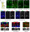
(A) Schematic drawing of muscles expressing Con in abdominal hemisegments. (B–E) Lateral views of abdominal hemisegments of stage 15–16 wild type (B), Df(3L)iroDFM3 (C), Con-GAL4::UAS-caupHA (D) and Con-GAL4::UAS-caupHA; UAS-rasV12 (E) embryos, stained with S59 antibody. Note the presence of an ectopic VA2 muscle (VA2*) in Df(3L)iroDFM3 (C), the absence of slou in VA2 when caup is ectopically expressed in this muscle (arrow, D, see also F-F″), and the failure of Caup to repress slou/S59 in VA2 muscle in the presence of the activated form of Ras, rasV12 (E). (F-G″) Lateral views of stage 15–16 Con-GAL4::UAS-caupHA; UAS-GFP (F-F″) and Con-GAL4::UAS-caupHA; UAS-slou (G-G″) embryos stained with the indicated antibodies. Note that co-expression of caup and slou in VA2 does not appreciably modify the morphology of the muscle (arrows in G-G″). As an internal control co-expression of UAS-caup and UAS-GFP still repressed endogenous slou and prevented the VA2 fate (F-F″). (H) Close-up of a lateral transverse promuscular cluster (outlined) in a stage 11 wild-type embryo showing co-expression of Caup (red) and Kr (blue) in all cells of the clusters. Note that the activation of the Ras/MAPK cascade (dpErk, green) only takes place at low levels in the segregating progenitor (yellow arrowhead) but not in the rest of the cluster. (I) Close-up of LT cluster in twist-GAL4; 24B-GAL4::UAS- rasV12 stage 11 embryo. Early activation of the Ras/MAPK pathway prevents the repression of slou by Caup in the LT cluster. (J) Close-up of the dorsal mesoderm of a mef2-GAL4::UAS-ara stage 15 embryo showing ectopic expression of slou in eve-expressing muscles.
Figure 8
Figure 8. Effect of the state of activation of the Ras/MAPK signalling cascade on the regulation of slou by Ara/Caup in LT and VA lineages.
In the wild-type LT3–4 promuscular cluster, where Ras/MAPK signalling is inactive, Caup represses slou since in embryos mutant for ara/caup (Df(3L)iroEGP6), the absence of Caup allows slou activation in this cluster and the consequent transformation of LT3–4 muscles to VA1–2 muscles. In the wild-type Caup is absent from the DER-dependent VA1–2 cluster that expresses slou. Ectopic expression of Caup in the VA1–2 lineages using Con-GAL4 (active after founder segregation when MAPK signalling is extinguished) represses slou in VA2. On the contrary, Con-GAL4 driven expression of Caup together with the activated form of Ras alleviates Caup-dependent slou repression in the VA2 muscle.

Similar articles

Cited by

References

    1. Bate M. The mesoderm and its derivatives; In: Martinez-Arias MBaA., editor. Cold Spring Harbor, New York: CHS Laboratory Press; 1993. pp. 1013–1090.
    1. Baylies MK, Bate M, Ruiz Gomez M. Myogenesis: a view from Drosophila. Cell. 1998;93:921–927. - PubMed
    1. Carmena A, Bate M, Jimenez F. Lethal of scute, a proneural gene, participates in the specification of muscle progenitors during Drosophila embryogenesis. Genes Dev. 1995;9:2373–2383. - PubMed
    1. Carmena A, Gisselbrecht S, Harrison J, Jimenez F, Michelson AM. Combinatorial signaling codes for the progressive determination of cell fates in the Drosophila embryonic mesoderm. Genes Dev. 1998;12:3910–3922. - PMC - PubMed
    1. Carmena A, Murugasu-Oei B, Menon D, Jimenez F, Chia W. Inscuteable and numb mediate asymmetric muscle progenitor cell divisions during Drosophila myogenesis. Genes Dev. 1998;12:304–315. - PMC - PubMed

Publication types

MeSH terms