. 2011 Jul 14:5:89.
doi: 10.3389/fnins.2011.00089. eCollection 2011.

A protocol for isolation and enriched monolayer cultivation of neural precursor cells from mouse dentate gyrus

Affiliations

A protocol for isolation and enriched monolayer cultivation of neural precursor cells from mouse dentate gyrus

Harish Babu et al. Front Neurosci. .

Abstract

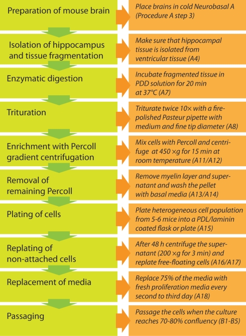
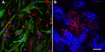
In vitro assays are valuable tools to study the characteristics of adult neural precursor cells under controlled conditions with a defined set of parameters. We here present a detailed protocol based on our previous original publication (Babu et al., 2007) to isolate neural precursor cells from the hippocampus of adult mice and maintain and propagate them as adherent monolayer cultures. The strategy is based on the use of Percoll density gradient centrifugation to enrich precursor cells from the micro-dissected dentate gyrus. Based on the expression of Nestin and Sox2, a culture-purity of more than 98% can be achieved. The cultures are expanded under serum-free conditions in Neurobasal A medium with addition of the mitogens Epidermal growth factor and Fibroblast growth factor 2 as well as the supplements Glutamax-1 and B27. Under differentiation conditions, the precursor cells reliably generate approximately 30% neurons with appropriate morphological, molecular, and electrophysiological characteristics that might reflect granule cell properties as their in vivo counterpart. We also highlight potential modifications to the protocol.

Keywords: adult neurogenesis; hippocampus; in vitro; precursor cell; progenitor cell.


Figures

Figure 1
Figure 1
Workflow diagram for the experimental procedure.
Figure 2
Figure 2
Highly enriched neural precursor cells propagated as adherent monolayer cultures (A–C). Adherent precursor cells cultures under routine proliferation conditions 1 day (A) and 5 days after seeding [(B) about 80% confluent; higher magnification in (C)]. (D–F) Upon differentiation with 2 days of 5 ng/ml FGF2 (D) and subsequent complete growth factor depletion (E,F), neural precursor cells change their morphology showing a progressively postmitotic and mature appearance of neuronal (arrows) and glial (asterisks) cells. Scale bar in (A): 100 μm for (A,B); 50 μm for (C–F).
Figure 3
Figure 3
In cultures of dentate gyrus neural precursor cells virtually all cells express cellular marker for proliferation, Ki67 (A), and for progenitor cells of the nervous system, such as Nestin (A–C), Sox2 (C) or vimentin (D). Many cells express also the radial glia marker BLBP (B) and Id1 (F), a dominant-negative bHLH transcriptional regulator expressed in a fraction of adult neural stem cells important for their self-renewal capacity. Only few cells express β3-tubulin (E) indicating neuronally committed cells. No cells were positive for S100β (F), a marker that labels mature astrocytes. Cells were grown until about 80% confluent (3–5 days) before fixing and staining. Blue stain in (A–F) is the nucleic dye DAPI. Scale bar in (A): 50 μm, in (A) and (B) for (A–F): 20 μm.
Figure 4
Figure 4
(A) Neural precursor cells upon 6 days of differentiation generate Map2ab (red) positive neurons and GFAP positive (green) glial cells. The nuclei are stained with DAPI (blue). (B) Ten days of differentiation generates O4 (red) positive oligodendrocytes. GFAP positive glial cells are depicted in blue.

Similar articles

Cited by

References

    1. Babu H., Cheung G., Kettenmann H., Palmer T. D., Kempermann G. (2007). Enriched monolayer precursor cell cultures from micro-dissected adult mouse dentate gyrus yield functional granule cell-like neurons. PLoS ONE 2, e388.10.1371/journal.pone.0000388 - DOI - PMC - PubMed
    1. Babu H., Ramirez-Rodriguez G., Fabel K., Bischofberger J., Kempermann G. (2009). Synaptic network activity induces neuronal differentiation of adult hippocampal precursor cells through BDNF signaling. Front. Neurosci. 3:49. - PMC - PubMed
    1. Ben Abdallah N., Slomianka L., Vyssotski A. L., Lipp H. P. (2010). Early age-related changes in adult hippocampal neurogenesis in C57 mice. Neurobiol. Aging 31, 151–16110.1016/j.neurobiolaging.2008.03.002 - DOI - PubMed
    1. Bez A., Corsini E., Curti D., Biggiogera M., Colombo A., Nicosia R. F., Pagano S. F., Parati E. A. (2003). Neurosphere and neurosphere-forming cells: morphological and ultrastructural characterization. Brain Res. 993, 18–2910.1016/j.brainres.2003.08.061 - DOI - PubMed
    1. Boku S., Nakagawa S., Masuda T., Nishikawa H., Kato A., Kitaichi Y., Inoue T., Koyama T. (2009). Glucocorticoids and lithium reciprocally regulate the proliferation of adult dentate gyrus-derived neural precursor cells through GSK-3beta and beta-catenin/TCF pathway. Neuropsychopharmacology 34, 805–81510.1038/npp.2008.198 - DOI - PubMed

LinkOut - more resources