. 2011 Apr 13:5:18.
doi: 10.3389/fnsys.2011.00018. eCollection 2011.

A high-light sensitivity optical neural silencer: development and application to optogenetic control of non-human primate cortex

Affiliations

A high-light sensitivity optical neural silencer: development and application to optogenetic control of non-human primate cortex

Xue Han et al. Front Syst Neurosci. .

Abstract

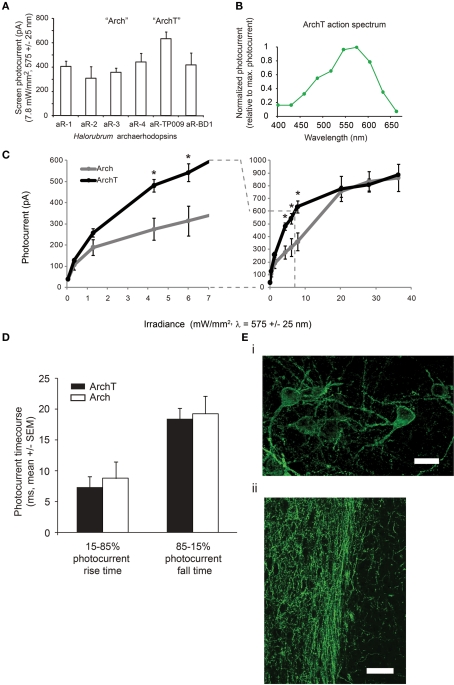
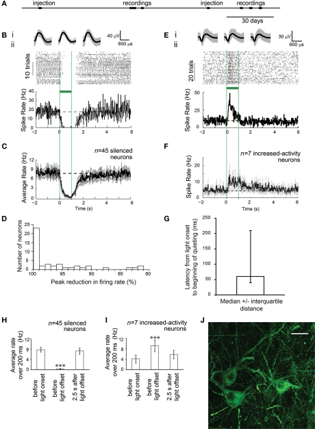
Technologies for silencing the electrical activity of genetically targeted neurons in the brain are important for assessing the contribution of specific cell types and pathways toward behaviors and pathologies. Recently we found that archaerhodopsin-3 from Halorubrum sodomense (Arch), a light-driven outward proton pump, when genetically expressed in neurons, enables them to be powerfully, transiently, and repeatedly silenced in response to pulses of light. Because of the impressive characteristics of Arch, we explored the optogenetic utility of opsins with high sequence homology to Arch, from archaea of the Halorubrum genus. We found that the archaerhodopsin from Halorubrum strain TP009, which we named ArchT, could mediate photocurrents of similar maximum amplitude to those of Arch (∼900 pA in vitro), but with a >3-fold improvement in light sensitivity over Arch, most notably in the optogenetic range of 1-10 mW/mm(2), equating to >2× increase in brain tissue volume addressed by a typical single optical fiber. Upon expression in mouse or rhesus macaque cortical neurons, ArchT expressed well on neuronal membranes, including excellent trafficking for long distances down neuronal axons. The high light sensitivity prompted us to explore ArchT use in the cortex of the rhesus macaque. Optical perturbation of ArchT-expressing neurons in the brain of an awake rhesus macaque resulted in a rapid and complete (∼100%) silencing of most recorded cells, with suppressed cells achieving a median firing rate of 0 spikes/s upon illumination. A small population of neurons showed increased firing rates at long latencies following the onset of light stimulation, suggesting the existence of a mechanism of network-level neural activity balancing. The powerful net suppression of activity suggests that ArchT silencing technology might be of great use not only in the causal analysis of neural circuits, but may have therapeutic applications.

Keywords: archaerhodopsin; channelrhodopsin; halorhodopsin; neural silencing; neurophysiology; non-human primate; optogenetics; systems neuroscience.


Figures

Figure 1
Figure 1
ArchT, a novel high-light sensitivity optical neural silencer. (A) Screen photocurrents generated by various opsins within the archaerhodopsin class, as assessed in neuron culture (n = 5–16 neurons for each bar), measured by whole-cell voltage clamp under standardized screening illumination conditions (7.8 mW/mm2, 575 ± 25 nm illumination). Abbreviations: aR-1, archaerhodopsin from Halorubrum strain aus-1; aR-2, archaerhodopsin from Halorubrum strain aus-2; aR-3, archaerhodopsin from Halorubrum sodomense (aka “Arch”); aR-4, archaerhodopsin from Halorubrum strain xz515; aR-TP009, archaerhodopsin from Halorubrum strain TP009 (aka “ArchT”); aR-BD1, archaerhodopsin from Halorubrum xingjianense. (B) Action spectrum of ArchT measured in cultured neurons by scanning illumination light wavelength through the visible spectrum (n = 8 neurons). (C) Photocurrents of ArchT and the previously published reagent Arch measured as a function of 575 ± 25 nm light irradiance (n = 4–16 neurons for each point); curves are Hill plots. *p < 0.05, t-test. (D) Rise times and fall times (measured from the time that the current takes to ramp up from 15 to 85% of its maximum value, and to decline by the same amount) for ArchT vs. Arch-mediated photocurrents in neurons. (Ei) Fluorescence image showing lentiviral ArchT-GFP expression in mouse motor cortex, ∼1 month after lentiviral injection. Scale bar, 15 μm. (Eii) Fluorescence image showing lentiviral ArchT-GFP expression in the axons of mouse infralimbic cortex neurons, here projecting into the nucleus accumbens shell, ∼1 month after lentiviral injection. Scale bar, 45 μm.
Figure 2
Figure 2
ArchT-mediated, ∼100% silencing of cortical neurons, in the awake primate brain. (A) Timeline of experiments for monkey A, indicating times of lentivirus (FCK-ArchT-GFP) injection and single-unit recording (data shown in B–G). (B) Neural activity in a representative silenced neuron in the primate brain before, during and after 1 s of green light illumination. (Bi) Action potential waveforms elicited before illumination (left), during illumination (middle), or after illumination (right); shown is the mean (black) as well as the overlay of raw waveforms (gray). (Bii) Neural activity, shown as a spike raster plot (top), and as a histogram of instantaneous firing rate averaged across trials (bottom; bin size 20 ms). (C) Histogram of instantaneous firing rate, averaged across all silenced single units recorded upon 1 s green light exposure, either using raw firing rate data (top), or using firing rate data normalized to baseline firing rate (bottom). Black line, mean; gray lines, mean ± standard error (SE); n = 45 silenced single units. (D) Histogram of peak percentage reductions in spike rate, for each individual silenced neuron. (E) Neural activity in a representative increased-activity neuron in the primate brain before, during and after 1 s of green light illumination. (Ei) Action potential waveforms elicited before illumination (left), during illumination (middle), or after illumination (right). (Eii) Neural activity, shown as a spike raster plot (top), and as a histogram of instantaneous firing rate averaged across trials (bottom; bin size 20 ms). (F) Histogram of instantaneous firing rate, averaged across all increased-activity single units recorded upon 200 ms green light exposure, either using raw firing rate data (top), or using firing rate data normalized to baseline firing rate (bottom). Black line, mean; gray lines, mean ± standard error (SE); n = 7 increased-activity single units. (G) Latencies from light onset to the beginning of quieting, for silenced neurons; plotted is median ± interquartile distance (n = 45 silenced neurons). (H) Summary histogram of all n = 45 silenced neurons, showing firing rates during 200 ms periods, either just before light onset, at the end of the light period (800–1000 ms), or at 2.5 s after light offset. ***p < 0.005, paired t-test, vs. baseline condition. (I) Summary histogram of all n = 7 increased-activity neurons, showing firing rates during 200 ms periods, either just before light onset, at the end of the light period (800–1000 ms), or at 2.5 s after light offset. (J) Fluorescence image showing lentiviral ArchT-GFP expression in monkey visual cortex, 1 month after lentiviral injection, in monkey B. Scale bar, 20 μm.

References

    1. Bainbridge J. W., Smith A. J., Barker S. S., Robbie S., Henderson R., Balaggan K., Viswanathan A., Holder G. E., Stockman A., Tyler N., Petersen-Jones S., Bhattacharya S. S., Thrasher A. J., Fitzke F. W., Carter B. J., Rubin G. S., Moore A. T., Ali R. R. (2008). Effect of gene therapy on visual function in Leber's congenital amaurosis. N. Engl. J. Med. 358, 2231–2239 - PubMed
    1. Nature Biotechnology, Editorial (2007). Retracing events. Nat. Biotechnol. 25, 949. - PubMed
    1. Boyden E. S., Zhang F., Bamberg E., Nagel G., Deisseroth K. (2005). Millisecond-timescale, genetically targeted optical control of neural activity. Nat. Neurosci. 8, 1263–1268 - PubMed
    1. Chow B. Y., Han X., Dobry A. S., Qian X., Chuong A. S., Li M., Henninger M. A., Belfort G. M., Lin Y., Monahan P. E., Boyden E. S. (2010). High-performance genetically targetable optical neural silencing by light-driven proton pumps. Nature 463, 98–10210.1038/nature08652 - DOI - PMC - PubMed
    1. Gradinaru V., Thompson K. R., Deisseroth K. (2008). eNpHR: a Natronomonas halorhodopsin enhanced for optogenetic applications. Brain Cell Biol. 36, 129–13910.1007/s11068-008-9027-6 - DOI - PMC - PubMed