. 2011 Jul 21:2:37.
doi: 10.3389/fphys.2011.00037. eCollection 2011.

Craniosynostosis of coronal suture in twist1 mice occurs through endochondral ossification recapitulating the physiological closure of posterior frontal suture

Affiliations

Craniosynostosis of coronal suture in twist1 mice occurs through endochondral ossification recapitulating the physiological closure of posterior frontal suture

Björn Behr et al. Front Physiol. .

Abstract

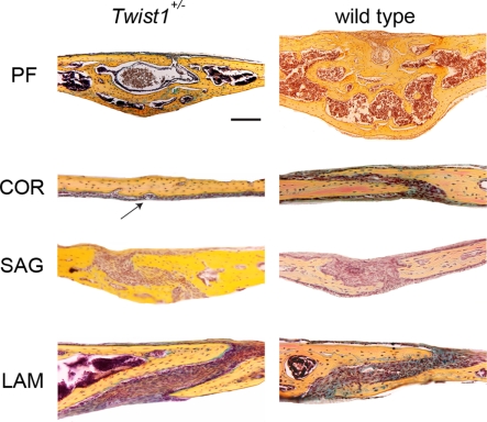
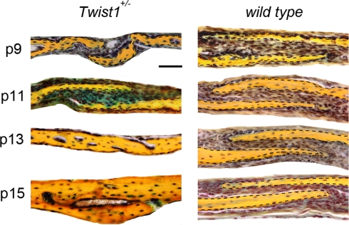
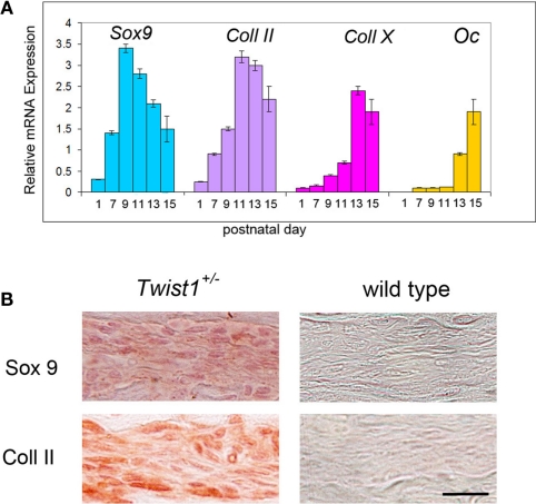
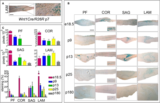
Craniosynostosis, the premature closure of cranial suture, is a pathologic condition that affects 1/2000 live births. Saethre-Chotzen syndrome is a genetic condition characterized by craniosynostosis. The Saethre-Chotzen syndrome, which is defined by loss-of-function mutations in the TWIST gene, is the second most prevalent craniosynostosis. Although much of the genetics and phenotypes in craniosynostosis syndromes is understood, less is known about the underlying ossification mechanism during suture closure. We have previously demonstrated that physiological closure of the posterior frontal suture occurs through endochondral ossification. Moreover, we revealed that antagonizing canonical Wnt-signaling in the sagittal suture leads to endochondral ossification of the suture mesenchyme and sagittal synostosis, presumably by inhibiting Twist1. Classic Saethre-Chotzen syndrome is characterized by coronal synostosis, and the haploinsufficient Twist1(+/-) mice represents a suitable model for studying this syndrome. Thus, we seeked to understand the underlying ossification process in coronal craniosynostosis in Twist1(+/-) mice. Our data indicate that coronal suture closure in Twist1(+/-) mice occurs between postnatal day 9 and 13 by endochondral ossification, as shown by histology, gene expression analysis, and immunohistochemistry. In conclusion, this study reveals that coronal craniosynostosis in Twist1(+/-) mice occurs through endochondral ossification. Moreover, it suggests that haploinsufficiency of Twist1 gene, a target of canonical Wnt-signaling, and inhibitor of chondrogenesis, mimics conditions of inactive canonical Wnt-signaling leading to craniosynostosis.

Keywords: canonical Wnt-signaling; cranial suture; craniosynostosis; endochondral ossification; twist haploinsufficiency.


Figures

Figure 1
Figure 1
Pentachrome staining of the four representative cranial sutures: posterior frontal (PF), coronal (COR), sagittal (SAG), and lambdoid (LAM) at postnatal day (p) 25. A complete closure of COR suture is observed in Twist1+/− mice, while the suture remains patent in wild-type mice. The arrow indicates the fused coronal suture in Twist1+/− mice. Scale bar: 100 μm.
Figure 2
Figure 2
Time course of COR suture closure in Twist1+/− mice. At day p11 pentachrome staining reveals the presence of cartilage (blue staining) between the bone plates of the COR suture of Twist1+/− mice, while no cartilage is present in wild-type mice. By day p13, pathological closure of COR suture is observed in Twist1+/− mice. Dashed lines indicate the bone plates. Scale bar: 50 μm.
Figure 3
Figure 3
Presence of specific chondrogenic markers in the differentiating COR suture mesenchyme. (A) Expression analysis of chondrogenic markers by qPCR showing a chondrogenic pattern of the differentiating COR suture mesenchyme in Twist1+/− mice. By postnatal day 15 upregulation of osteocalcin (Oc) is observed, as result of the COR suture closure. (B) Immunohistochemistry for Sox9 and Col II confirm cartilage tissue formation in the COR suture of Twist1+/− at day p11. Scale bar: 10 μm Expression analysis of chondrogenic markers by qPCR showing a chondrogenic pattern of the differentiating COR suture mesenchyme.
Figure 4
Figure 4
Activation of canonical Wnt-signaling in embryonic and postnatal cranial sutures. (A) Xgal staining of LAM suture harvested from Wnt1Cre/R26R mouse. Xgal staining indicates the neural-crest origin of the LAM suture mesenchyme. The boxed area is enlarged to the right. (B) Xgal staining of PF, COR SAG, and LAM suture mesenchymes in Axin2+/− mice from e18.5 to p180. Scale bar: 100 μm. Inserts represent zoomed areas of the coronal sutures. Dashed lines indicate the bone plates. (C) Quantification of Xgal staining of different cranial suture mesenchymes in Axin2+/− mice at day p25 and p180.

References

    1. Behr B., Longaker M. T., Quarto N. (2010). Differential activation of canonical Wnt signaling determines cranial sutures fate: a novel mechanism for sagittal suture craniosynostosis. Dev. Biol. 344, 922–94010.1016/j.ydbio.2010.06.009 - DOI - PubMed
    1. Bellus G. A., Gaudenz K., Zackai E. H., Clarke L. A., Szabo J., Francomano C. A., Muenke M. (1996). Identical mutations in three different fibroblast growth factor receptor genes in autosomal dominant craniosynostosis syndromes. Nat. Genet. 14, 174–17610.1038/ng1096-174 - DOI - PubMed
    1. Bialek P., Kern B., Yang X., Schrock M., Sosic D., Hong N., Wu H., Yu K., Ornitz D. M., Olson E. N., Justice M. J., Karsenty G. (2004). A twist code determines the onset of osteoblast differentiation. Dev. Cell 6, 423–43510.1016/S1534-5807(04)00058-9 - DOI - PubMed
    1. Bourgeois P., Bolcato-Bellemin A. L., Danse J. M., Bloch-Zupan A., Yoshiba K., Stoetzel C., Perrin-Schmitt F. (1998). The variable expressivity and incomplete penetrance of the twist-null heterozygous mouse phenotype resemble those of human Saethre-Chotzen syndrome. Hum. Mol. Genet. 7, 945–95710.1093/hmg/7.6.945 - DOI - PubMed
    1. Carver E. A., Oram K. F., Gridley T. (2002). Craniosynostosis in Twist heterozygous mice: a model for Saethre-Chotzen syndrome. Anat. Rec. 268, 90–9210.1002/ar.10124 - DOI - PubMed

LinkOut - more resources