

Experimental gastric carcinogenesis in Cebus apella nonhuman primates
- PMID: 21811552
- PMCID: PMC3140998
- DOI: 10.1371/journal.pone.0021988
Experimental gastric carcinogenesis in Cebus apella nonhuman primates
Abstract
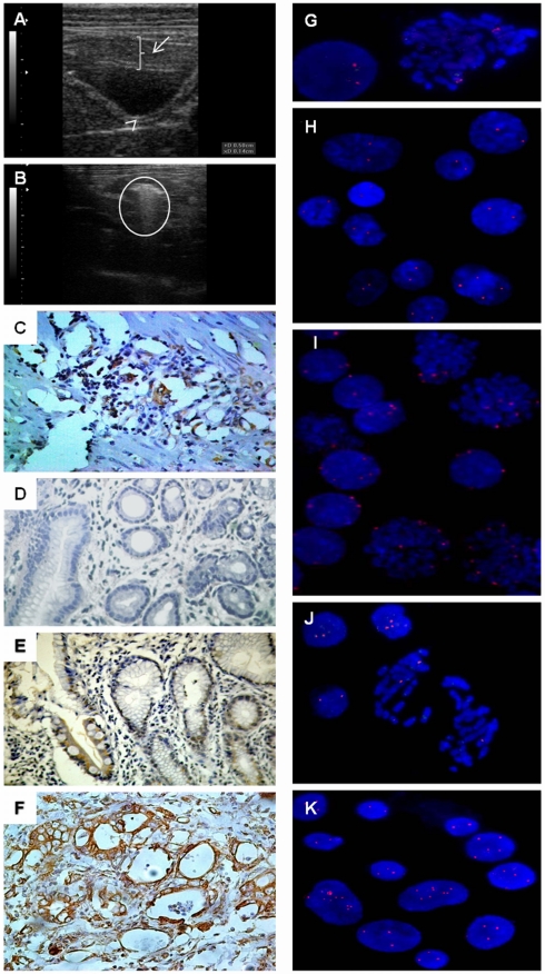
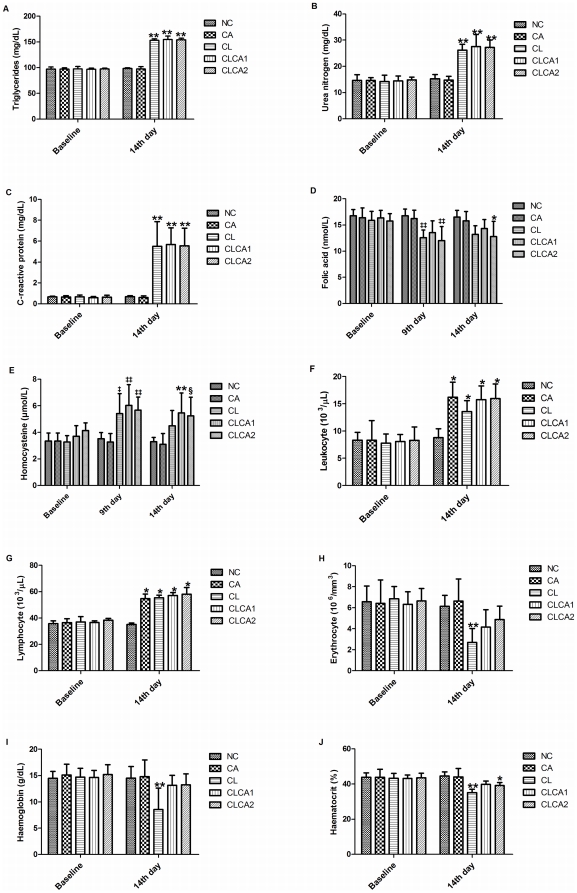
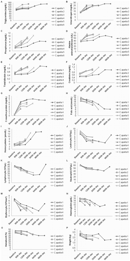
The evolution of gastric carcinogenesis remains largely unknown. We established two gastric carcinogenesis models in New-World nonhuman primates. In the first model, ACP03 gastric cancer cell line was inoculated in 18 animals. In the second model, we treated 6 animals with N-methyl-nitrosourea (MNU). Animals with gastric cancer were also treated with Canova immunomodulator. Clinical, hematologic, and biochemical, including C-reactive protein, folic acid, and homocysteine, analyses were performed in this study. MYC expression and copy number was also evaluated. We observed that all animals inoculated with ACP03 developed gastric cancer on the 9(th) day though on the 14(th) day presented total tumor remission. In the second model, all animals developed pre-neoplastic lesions and five died of drug intoxication before the development of cancer. The last surviving MNU-treated animal developed intestinal-type gastric adenocarcinoma observed by endoscopy on the 940(th) day. The level of C-reactive protein level and homocysteine concentration increased while the level of folic acid decreased with the presence of tumors in ACP03-inoculated animals and MNU treatment. ACP03 inoculation also led to anemia and leukocytosis. The hematologic and biochemical results corroborate those observed in patients with gastric cancer, supporting that our in vivo models are potentially useful to study this neoplasia. In cell line inoculated animals, we detected MYC immunoreactivity, mRNA overexpression, and amplification, as previously observed in vitro. In MNU-treated animals, mRNA expression and MYC copy number increased during the sequential steps of intestinal-type gastric carcinogenesis and immunoreactivity was only observed in intestinal metaplasia and gastric cancer. Thus, MYC deregulation supports the gastric carcinogenesis process. Canova immunomodulator restored several hematologic measurements and therefore, can be applied during/after chemotherapy to increase the tolerability and duration of anticancer treatments.
Conflict of interest statement
Figures
Similar articles
-
Evaluation of the immunological cellular response of Cebus apella exposed to the carcinogen N-methyl-N-nitrosourea and treated with CANOVA®.In Vivo. 2014 Sep-Oct;28(5):837-41. In Vivo. 2014. PMID: 25189897
-
Deregulation of the SRC Family Tyrosine Kinases in Gastric Carcinogenesis in Non-human Primates.Anticancer Res. 2018 Nov;38(11):6317-6320. doi: 10.21873/anticanres.12988. Anticancer Res. 2018. PMID: 30396952
-
MYC, FBXW7 and TP53 copy number variation and expression in gastric cancer.BMC Gastroenterol. 2013 Sep 23;13:141. doi: 10.1186/1471-230X-13-141. BMC Gastroenterol. 2013. PMID: 24053468 Free PMC article.
-
The Complex Network between MYC Oncogene and microRNAs in Gastric Cancer: An Overview.Int J Mol Sci. 2020 Mar 5;21(5):1782. doi: 10.3390/ijms21051782. Int J Mol Sci. 2020. PMID: 32150871 Free PMC article. Review.
-
MYC and gastric adenocarcinoma carcinogenesis.World J Gastroenterol. 2008 Oct 21;14(39):5962-8. doi: 10.3748/wjg.14.5962. World J Gastroenterol. 2008. PMID: 18932273 Free PMC article. Review.
Cited by
-
What gastric cancer proteomic studies show about gastric carcinogenesis?Tumour Biol. 2016 Aug;37(8):9991-10010. doi: 10.1007/s13277-016-5043-9. Epub 2016 Apr 28. Tumour Biol. 2016. PMID: 27126070 Review.
-
Gastric cancer and gene copy number variation: emerging cancer drivers for targeted therapy.Oncogene. 2016 Mar 24;35(12):1475-82. doi: 10.1038/onc.2015.209. Epub 2015 Jun 15. Oncogene. 2016. PMID: 26073079 Review.
-
CDKN1A histone acetylation and gene expression relationship in gastric adenocarcinomas.Clin Exp Med. 2017 Feb;17(1):121-129. doi: 10.1007/s10238-015-0400-3. Epub 2015 Nov 14. Clin Exp Med. 2017. PMID: 26567008
-
Deregulated Expression of SRC, LYN and CKB Kinases by DNA Methylation and Its Potential Role in Gastric Cancer Invasiveness and Metastasis.PLoS One. 2015 Oct 13;10(10):e0140492. doi: 10.1371/journal.pone.0140492. eCollection 2015. PLoS One. 2015. PMID: 26460485 Free PMC article.
-
hTERT, MYC and TP53 deregulation in gastric preneoplastic lesions.BMC Gastroenterol. 2012 Jul 6;12:85. doi: 10.1186/1471-230X-12-85. BMC Gastroenterol. 2012. PMID: 22768805 Free PMC article.
References
-
- Parkin DM, Bray F, Ferlay J, Pisani P. Global cancer statistics, 2002. CA Cancer J Clin. 2005;55:74–108. - PubMed
-
- Prater MR, Zimmerman KL, Ward DL, Holladay SD. Reduced birth defects caused by maternal immune stimulation in methylnitrosourea-exposed mice: association with placental improvement. Birth Defects Res A Clin Mol Teratol. 2004;70:862–869. - PubMed
Publication types
MeSH terms
Substances
LinkOut - more resources
Full Text Sources
Medical
Research Materials
