. 2011;6(7):e22435.
doi: 10.1371/journal.pone.0022435. Epub 2011 Jul 21.

Cable pili and the associated 22 kDa adhesin contribute to Burkholderia cenocepacia persistence in vivo

Affiliations

Cable pili and the associated 22 kDa adhesin contribute to Burkholderia cenocepacia persistence in vivo

Joanna B Goldberg et al. PLoS One. 2011.

Abstract

Background: Infection by Burkholderia cenocepacia in cystic fibrosis (CF) patients is associated with poor clinical prognosis. Previously, we demonstrated that one of the highly transmissible strains, BC7, expresses cable pili and the associated 22 kDa adhesin, both of which contribute to BC7 binding to airway epithelial cells. However, the contribution of these factors to induce inflammation and bacterial persistence in vivo is not known.

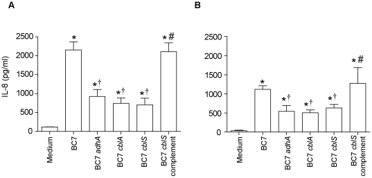
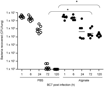
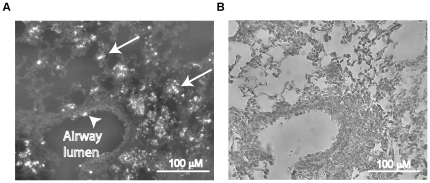
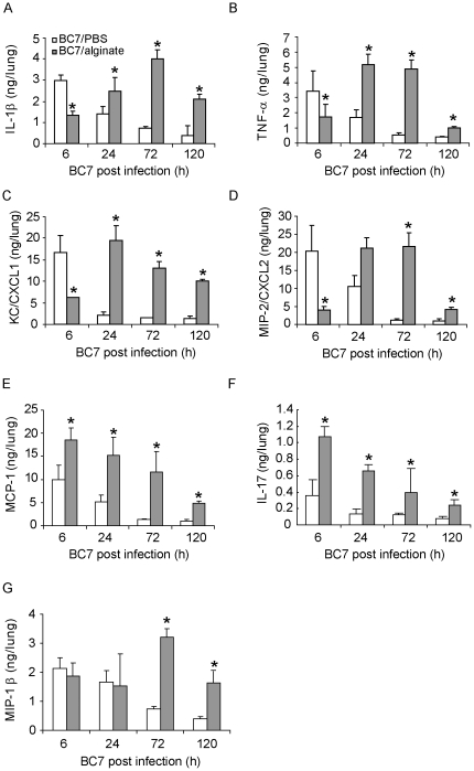
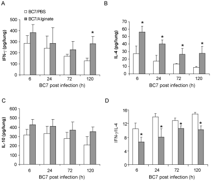
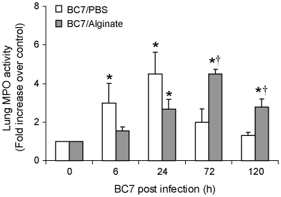
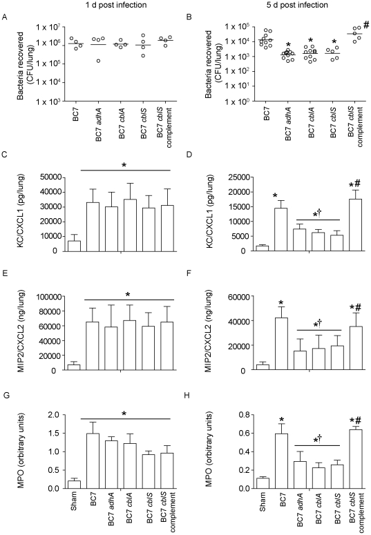
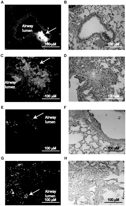
Methodology/principal findings: Wild-type BC7 stimulated higher IL-8 responses than the BC7 cbl and BC7 adhA mutants in both CF and normal bronchial epithelial cells. To determine the role of cable pili and the associated adhesin, we characterized a mouse model of B. cenocepacia, where BC7 are suspended in Pseudomonas aeruginosa alginate. C57BL/6 mice were infected intratracheally with wild-type BC7 suspended in either alginate or PBS and were monitored for lung bacterial load and inflammation. Mice infected with BC7 suspended in PBS completely cleared the bacteria by 3 days and resolved the inflammation. In contrast, mice infected with BC7 suspended in alginate showed persistence of bacteria and moderate lung inflammation up to 5 days post-infection. Using this model, mice infected with the BC7 cbl and BC7 adhA mutants showed lower bacterial loads and mild inflammation compared to mice infected with wild-type BC7. Complementation of the BC7 cblS mutation in trans restored the capacity of this strain to persist in vivo. Immunolocalization of bacteria revealed wild-type BC7 in both airway lumen and alveoli, while the BC7 cbl and BC7 adhA mutants were found mainly in airway lumen and peribronchiolar region.

Conclusions and significance: B. cenocepacia suspended in alginate can be used to determine the capacity of bacteria to persist and cause lung inflammation in normal mice. Both cable pili and adhesin contribute to BC7-stimulated IL-8 response in vitro, and BC7 persistence and resultant inflammation in vivo.


Conflict of interest statement

Competing Interests: The authors have declared that no competing interests exist.

Figures

Figure 1
Figure 1. Stimulation of IL-8 response by B. cenocepacia strains in airway epithelial cells.
IB3 (A) or BEAS2B (B) cells were treated with media or media containing BC7, BC7 adhA, BC7 cblA, BC7 cblS, or the BC7 cblS complemented strain (BC7 cblS complement), and IL-8 was determined by ELISA. Data represents mean ± SEM calculated from three independent experiments carried out in triplicates. (*different from medium control, † different from BC7, #different from BC7 mutants p≤0.05, ANOVA).
Figure 2
Figure 2. Lung bacterial load in C57BL/6 mice infected with BC7/PBS or BC7/alginate.
Mice infected with BC7/PBS or BC7/alginate were sacrificed at pre-determined times, and ten-fold serial dilutions of lung homogenates were plated on BCSA to determine bacterial load. Data points represent individual mice and bars represent geometric mean calculated from three independent experiments with a total of 6 - 7 mice per group (*different from BC7/PBS group at respective time points, p≤0.05, ANOVA on Ranks).
Figure 3
Figure 3. Immunolocalization of B. cenocepacia in mice infected with BC7/alginate.
Mice were infected with BC7/alginate and sacrificed one day after infection. Lungs were fixed and embedded in paraffin. Lung sections were deparaffinized and incubated with antibody to B. cenocepacia. Bound antibody was detected by anti-rabbit IgG conjugated with Alexafluor 598 and sections were visualized under fluorescence microscope. A, lung section showing bacteria in both conducting (arrowhead) and respiratory zone (arrow). B, bright field image of A. Images are representative of two independent experiments.
Figure 4
Figure 4. Cytokine levels in lungs of mice infected with BC7.
Mice infected with BC7/PBS or BC7/alginate were sacrificed at specified times, and the cytokine levels in lung homogenates were measured by Bio-Plex multiplex immunoassay. A, IL-1β; B, TNF-α; C, KC/CXCL1; D, MIP-2/CXCL2; E, MCP-1; F, IL-17; and G, MIP-1β. Data represents mean and SEM calculated from three independent experiments with a total of 6 - 7 mice per group (*different from BC7/PBS group at respective time points, p≤0.05, ANOVA).
Figure 5
Figure 5. Th1 and Th2 cytokine levels in mice infected with BC7.
Mice infected with BC7/PBS or BC7/alginate were sacrificed at specified times and levels of IFN-γ (A), IL-4 (B), and IL-10 (C) in lung homogenates were measured by Bio-Plex multiple immunassay. D, ratio of IFN-γ to IL-4. Data represents mean and SEM calculated from three independent experiments with a total of 6 - 7 mice per group (*different from BC7/PBS group at respective time points, p≤ANOVA).
Figure 6
Figure 6. Lung MPO activity in BC7 infected C57BL/6 mice.
Mice infected with BC7/PBS or BC7/alginate were sacrificed at specified times and MPO activity was determined in the lung homogenates. Data represents mean and SEM calculated from three independent experiments with a total of 6-7 mice per group (*different from respective uninfected group p≤0.05, †different from BC7/PBS group at respective time points p≤0.05, ANOVA).
Figure 7
Figure 7. Lung inflammation in C57BL/6 mice infected with BC7/PBS or BC7/alginate.
Paraffin lung sections from mice infected with BC7/PBS (A, C, E and G) or BC7/alginate (B, D, F and H) were stained with H&E. A and B, 6 h post-infection; C and D, 1 d post-infection; E and F, 3 d post-infection; and G and H, 5 d post-infection, respectively. Insets in A, C, D, F and H represent magnified view of areas marked in respective panels and show infiltrated neutrophils. Images are representative of 3 individual animals at each time point.
Figure 8
Figure 8. Bacterial load and chemokine responses in mice infected with B. cenocepacia strains.
Mice were infected with wild-type BC7, the BC7 adhA, BC7 cblA, BC7 cblS mutants, or the BC7 cblS complement, suspended in alginate, and sacrificed at 1 or 5 d post-infection. Lung homogenates were used to determine bacterial load (A and B), KC (C and D), MIP-2 (E and F) and MPO activity (G and H). Data in represents either median with range (A) or mean with SEM (B–D) determined from two independent experiments with a total of 5 mice per group (*different from wild-type BC7, † different from BC7, #different from BC7 mutants p≤0.05, ANOVA on Ranks or ANOVA).
Figure 9
Figure 9. Immunolocalization of bacteria in mice infected with BC7 mutants or BC7 cblS complement.
Mice were infected with the BC7 adhA, BC7 cblA, or BC7 cblS mutants or the BC7 cblS complement suspended in alginate and sacrificed at 1 d post-infection. Paraffin lung sections from these mice were deparaffinized and incubated with antibody to B. cenocepacia. Bound antibody was detected by second antibody conjugated with AlexaFluor 488 and sections were counterstained with hematoxylin. Sections were viewed under fluorescence microscope. A, C, E and G shows bacteria in lung sections from mice infected with BC7 adhA, BC7 cblA, BC7 cblS, and the BC7 cblS complement, respectively. B, D, F and H are bright field images of A, C, E and G respectively. Arrows point to bacteria in the lung. Images are representative of three mice per group.

Similar articles

Cited by

References

    1. Mahenthiralingam E, Baldwin A, Dowson CG. Burkholderia cepacia complex bacteria: opportunistic pathogens with important natural biology. J Appl Microbiol. 2008;104:1539–1551. - PubMed
    1. Vanlaere E, Lipuma JJ, Baldwin A, Henry D, De Brandt E, et al. Burkholderia latens sp. nov., Burkholderia diffusa sp. nov., Burkholderia arboris sp. nov., Burkholderia seminalis sp. nov. and Burkholderia metallica sp. nov., novel species within the Burkholderia cepacia complex. Int J Syst Evol Microbiol. 2008;58:1580–1590. - PubMed
    1. Mahenthiralingam E, Urban TA, Goldberg JB. The multifarious, multireplicon Burkholderia cepacia complex. Nat Rev Microbiol. 2005;3:144–156. - PubMed
    1. Isles A, Maclusky I, Corey M, Gold R, Prober C, et al. Pseudomonas cepacia infection in cystic fibrosis: an emerging problem. J Pediatr. 1984;104:206–210. - PubMed
    1. Sajjan US, Corey M, Karmali MA, Forstner JF. Binding of Pseudomonas cepacia to normal human intestinal mucin and respiratory mucin from patients with cystic fibrosis. J Clin Invest. 1992;89:648–656. - PMC - PubMed

Publication types

MeSH terms