. 2011 Jul;5(7):e1237.
doi: 10.1371/journal.pntd.0001237. Epub 2011 Jul 19.

Mycolactone diffuses into the peripheral blood of Buruli ulcer patients--implications for diagnosis and disease monitoring

Affiliations

Mycolactone diffuses into the peripheral blood of Buruli ulcer patients--implications for diagnosis and disease monitoring

Fred S Sarfo et al. PLoS Negl Trop Dis. 2011 Jul.

Abstract

Background: Mycobacterium ulcerans, the causative agent of Buruli ulcer (BU), is unique among human pathogens in its capacity to produce a polyketide-derived macrolide called mycolactone, making this molecule an attractive candidate target for diagnosis and disease monitoring. Whether mycolactone diffuses from ulcerated lesions in clinically accessible samples and is modulated by antibiotic therapy remained to be established.

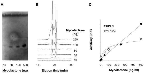
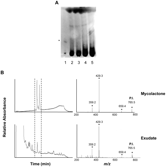
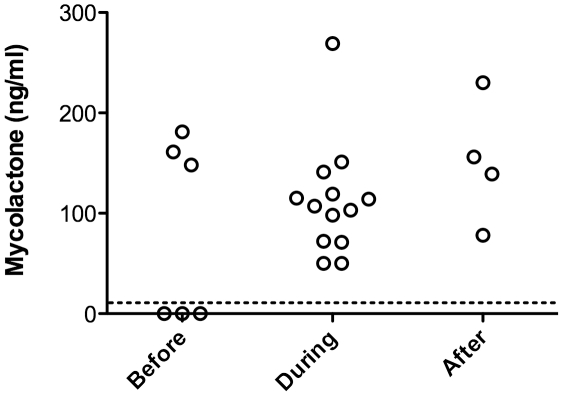
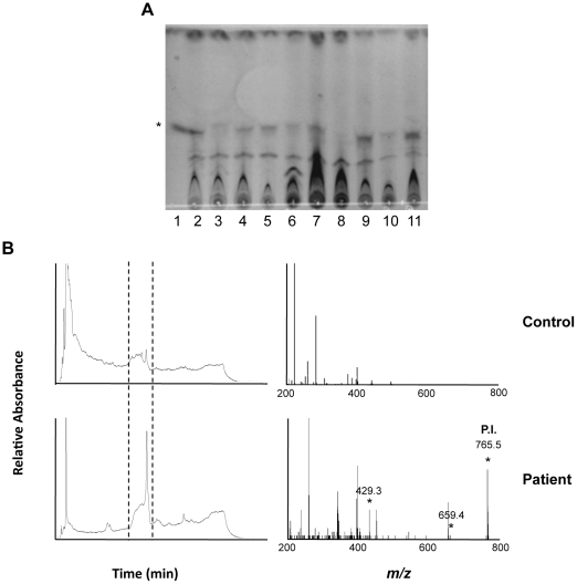
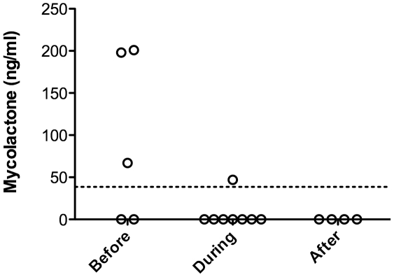
Methodology/principal finding: Peripheral blood and ulcer exudates were sampled from patients at various stages of antibiotic therapy in Ghana and Ivory Coast. Total lipids were extracted from serum, white cell pellets and ulcer exudates with organic solvents. The presence of mycolactone in these extracts was then analyzed by a recently published, field-friendly method using thin layer chromatography and fluorescence detection. This approach did not allow us to detect mycolactone accurately, because of a high background due to co-extracted human lipids. We thus used a previously established approach based on high performance liquid chromatography coupled to mass spectrometry. By this means, we could identify structurally intact mycolactone in ulcer exudates and serum of patients, and evaluate the impact of antibiotic treatment on the concentration of mycolactone.

Conclusions/significance: Our study provides the proof of concept that assays based on mycolactone detection in serum and ulcer exudates can form the basis of BU diagnostic tests. However, the identification of mycolactone required a technology that is not compatible with field conditions and point-of-care assays for mycolactone detection remain to be worked out. Notably, we found mycolactone in ulcer exudates harvested at the end of antibiotic therapy, suggesting that the toxin is eliminated by BU patients at a slow rate. Our results also indicated that mycolactone titres in the serum may reflect a positive response to antibiotics, a possibility that it will be interesting to examine further through longitudinal studies.


Conflict of interest statement

The authors have declared that no competing interests exist.

Figures

Figure 1
Figure 1. Comparison of the performances of the TLC-Fluo and HPLC approaches for mycolactone quantitative detection.
A representative picture of the fluorescent signals and elution peaks obtained for mycolactone (10–500 ng) detection by TLC-Fluo (A) and HPLC (B) are shown, with the corresponding standard curves (C). Arbitrary units correspond to fluorescence intensity of the mycolactone band (TLC-Fluo) and area of the mycolactone elution peak (HPLC). Similar results were obtained in at least three independent experiments.
Figure 2
Figure 2. Structurally intact mycolactone is detected in ulcer exudates of BU patients.
(A) Example of the fluorescence signals given by ulcer exudates (lanes 2–5), compared to pure mycolactone (lane 1) following analysis by TLC-Fluo. The band corresponding to mycolactone is designated by an asterisk. (B) HPLC elution profiles are shown for reference mycolactone (100 ng) and for one representative ulcer exudate among the 20 positive ones. The corresponding MS/MS spectra are presented, with mycolactone parent ion (P.I.) and products designated by asterisks. Similar MS/MS spectra were obtained from HPLC elution peaks collected from three positive patients.
Figure 3
Figure 3. Mycolactone presence is maintained in ulcer exudates during antibiotic therapy.
Mean concentration of mycolactone in ulcer exudates harvested before (0 week), during (2 to 8 weeks of treatment), or after completion of the 8 week antibiotic treatment. Dashed horizontal line indicates detection threshold.
Figure 4
Figure 4. Structurally intact mycolactone is detected in serum samples of BU patients.
(A) Example of the fluorescence signals given by serum samples (lanes 2–9), following lipid extraction and analysis by TLC-Fluo. Controls include 1 µg pure mycolactone (lane 1), lipids extracted from a negative serum (lane 10), and then spiked with 1 µg mycolactone (lane 11). The band corresponding to mycolactone is designated by an asterisk. (B) Representative HPLC elution profiles of lipids extracted from serum samples are shown for one healthy control out of 5, and one BU patient among the 4 positive ones. The corresponding MS/MS spectra show the presence of the parent and product ions of mycolactone in this positive sample. Similar results were obtained in a second one.
Figure 5
Figure 5. Mycolactone concentration in the serum of BU patients during antibiotic therapy.
Mean concentration of mycolactone in serum samples collected before (0 week), during (2 to 8 weeks of treatment), or after completion of the 8 week antibiotic treatment. Dashed horizontal line indicates detection threshold.

References

    1. Silva MT, Portaels F, Pedrosa J. Pathogenetic mechanisms of the intracellular parasite Mycobacterium ulcerans leading to Buruli ulcer. Lancet Infect Dis. 2009;9:699–710. - PubMed
    1. Walsh DS, Portaels F, Meyers WM. Recent advances in leprosy and Buruli ulcer (Mycobacterium ulcerans infection). Curr Opin Infect Dis. 2010;23:445–455. - PubMed
    1. Etuaful S, Carbonnelle B, Grosset J, Lucas S, Horsfield C, et al. Efficacy of the combination rifampin-streptomycin in preventing growth of Mycobacterium ulcerans in early lesions of Buruli ulcer in humans. Antimicrob Agents Chemother. 2005;49:3182–3186. - PMC - PubMed
    1. Chauty A, Ardant MF, Adeye A, Euverte H, Guedenon A, et al. Promising clinical efficacy of streptomycin-rifampin combination for treatment of buruli ulcer (Mycobacterium ulcerans disease). Antimicrob Agents Chemother. 2007;51:4029–4035. - PMC - PubMed
    1. Johnson PD. Should antibiotics be given for Buruli ulcer? Lancet. 2010;375:618–619. - PubMed

Publication types

MeSH terms