

Optical coherence tomography findings in idiopathic macular holes
- PMID: 21811669
- PMCID: PMC3147006
- DOI: 10.1155/2011/928205
Optical coherence tomography findings in idiopathic macular holes
Abstract
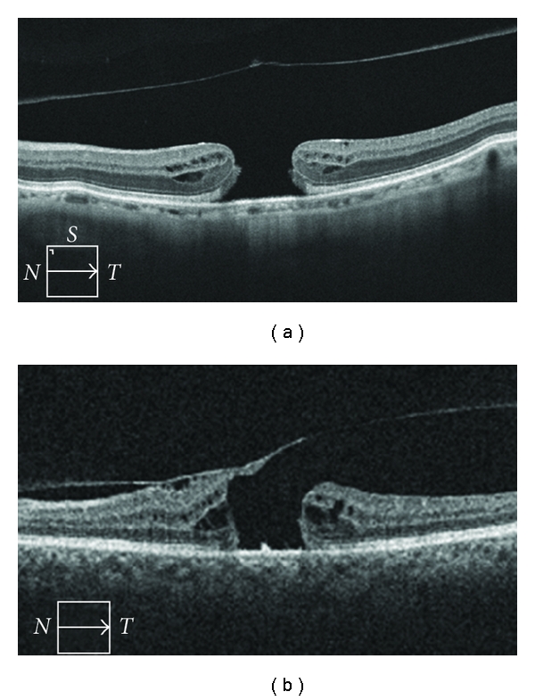
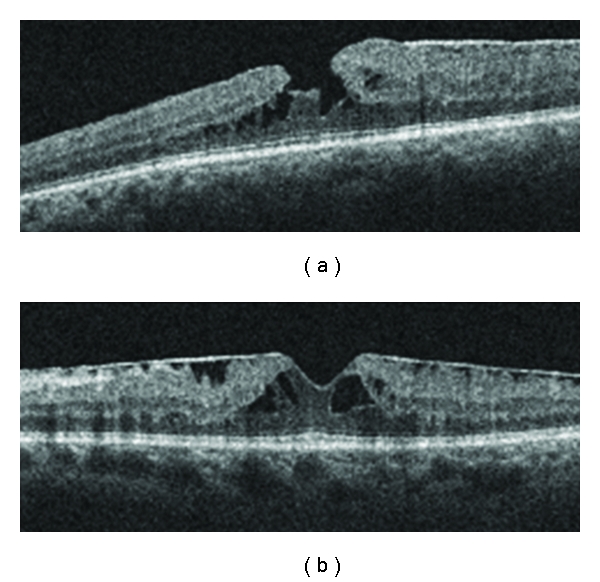
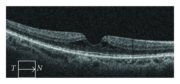
Purpose. To describe the characteristics of idiopathic macular holes (MH) on optical coherence tomography (OCT) and correlate OCT with clinical assessment. Design. Cross-sectional chart review and OCT assessment. Participants. Sixty-seven eyes with a clinically diagnosed idiopathic MH with available OCT data. Methods. A retrospective chart review and OCT assessment. Results. Based on OCT grading, 40 eyes had a full-thickness macular hole (FTMH) and 21 eyes had a lamellar macular hole (LMH). Clinical exam and OCT assessment agreed in 53 (87%) eyes when assessing the extent of MH. Six eyes (14.6%) in the FTMH group, and 3 eyes in the LMH group (14.3%) had persistent vitreomacular traction. Thirty-seven eyes (92.5%) in the FTMH group and 11 eyes (52.4%) in the LMH group had associated intraretinal cysts. Two eyes (5.0%) in the FTMH group and zero eyes in the LMH group had subretinal fluid. Intraretinal cysts were found to be more frequently associated with FTMH than with LMH (P < 0.001). Conclusion. This paper described OCT findings in a group of patients with clinically diagnosed MH. A high level of correlation between clinical assessment and OCT findings of LMH and FTMH was observed, and intraretinal cysts were often present in FTMH.
Figures
Similar articles
-
Risk Factors for Progression of Vitreomacular Traction to Macular Hole.J Vitreoretin Dis. 2024 Jul 27;8(5):524-532. doi: 10.1177/24741264241264937. eCollection 2024 Sep-Oct. J Vitreoretin Dis. 2024. PMID: 39381333 Free PMC article.
-
[Morphology of the vitreoretinal interface in fellow eyes of patients with full thickness macular holes].Ophthalmologe. 2018 Dec;115(12):1050-1055. doi: 10.1007/s00347-017-0614-8. Ophthalmologe. 2018. PMID: 29138978 German.
-
Hyperreflective Stress Lines and Macular Holes.Invest Ophthalmol Vis Sci. 2020 Apr 9;61(4):50. doi: 10.1167/iovs.61.4.50. Invest Ophthalmol Vis Sci. 2020. PMID: 32347919 Free PMC article.
-
Primary Lamellar Macular Holes: To Vit or Not to Vit.J Clin Med. 2022 Aug 28;11(17):5046. doi: 10.3390/jcm11175046. J Clin Med. 2022. PMID: 36078977 Free PMC article. Review.
-
[Lamellar macular holes with hyporeflective epiretinal proliferation : OCT diagnostics and clinical course].Ophthalmologe. 2017 Dec;114(12):1100-1109. doi: 10.1007/s00347-017-0597-5. Ophthalmologe. 2017. PMID: 29110126 Review. German.
Cited by
-
Clinical application of ocular imaging.Optom Vis Sci. 2012 May;89(5):E543-53. doi: 10.1097/OPX.0b013e31824f164d. Optom Vis Sci. 2012. PMID: 22488266 Free PMC article. Review.
-
Correlations between intraretinal cystoid cavities and pre- and postoperative characteristics of eyes after closure of idiopathic macular hole.Sci Rep. 2020 Feb 11;10(1):2310. doi: 10.1038/s41598-020-59295-7. Sci Rep. 2020. PMID: 32047222 Free PMC article.
-
Vitreoretinal Interface Characteristics in Eyes with Idiopathic Macular Holes: Qualitative and Quantitative Analysis.Turk J Ophthalmol. 2018 Apr;48(2):70-74. doi: 10.4274/tjo.23327. Epub 2018 Apr 25. Turk J Ophthalmol. 2018. PMID: 29755819 Free PMC article.
-
Bilateral simultaneous stage 1 macular hole.Ophthalmic Surg Lasers Imaging. 2012 Sep 27;43 Online:e99-e103. doi: 10.3928/15428877-20120920-04. Ophthalmic Surg Lasers Imaging. 2012. PMID: 23053850 Free PMC article.
-
Comparative study of combined vitrectomy with phacoemulsification versus vitrectomy alone for primary full-thickness macular hole repair.BMC Ophthalmol. 2021 Apr 10;21(1):174. doi: 10.1186/s12886-021-01918-2. BMC Ophthalmol. 2021. PMID: 33838649 Free PMC article.
References
-
- Johnson RN, Gass JD. Idiopathic macular holes. Observation, stages of formation, and implications for surgical intervention. Ophthalmology. 1988;95(7):917–924. - PubMed
-
- Gass JD. Reappraisal of biomicroscopic classification of stages of development of a macular hole. American Journal of Ophthalmology. 1995;119(6):752–759. - PubMed
-
- Martinez J, Smiddy WE, Kim J, Gass JD. Differentiating macular holes from macular pseudoholes. American Journal of Ophthalmology. 1994;117(6):762–767. - PubMed
-
- Azzolini C, Patelli F, Brancato R. Correlation between optical coherence tomography data and biomicroscopic interpretation of idiopathic macular hole. American Journal of Ophthalmology. 2001;132(3):348–355. - PubMed
LinkOut - more resources
Full Text Sources
