. 2011 Sep 15;118(11):3028-38.
doi: 10.1182/blood-2011-04-348839. Epub 2011 Aug 2.

TLR7 enables cross-presentation by multiple dendritic cell subsets through a type I IFN-dependent pathway

Affiliations

TLR7 enables cross-presentation by multiple dendritic cell subsets through a type I IFN-dependent pathway

Jason Z Oh et al. Blood. .

Abstract

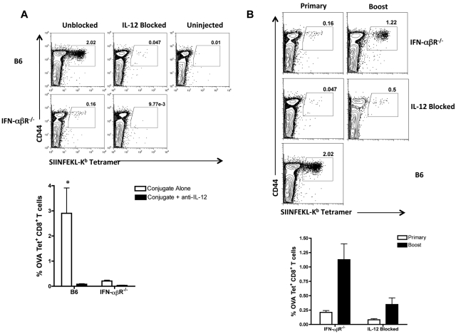
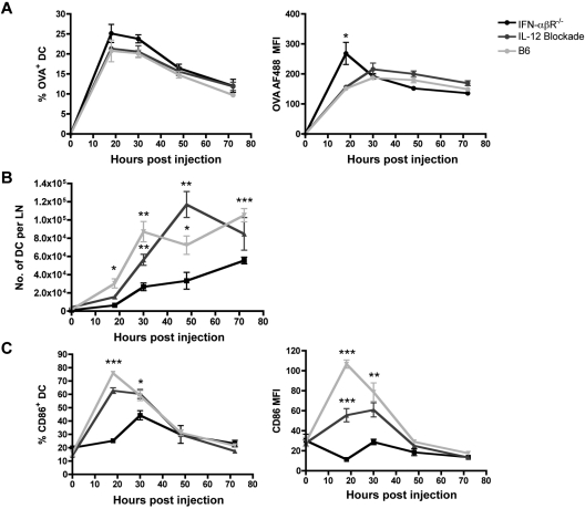
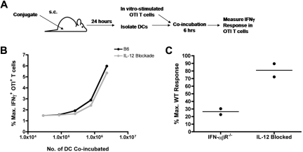
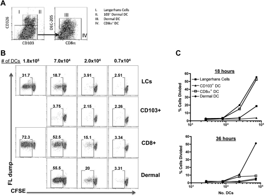
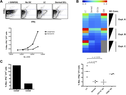
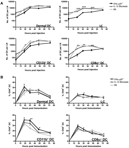
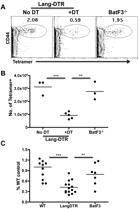
Conjugation of TLR agonists to protein or peptide antigens has been demonstrated in many studies to be an effective vaccine formula in inducing cellular immunity. However, the molecular and cellular mediators involved in TLR-induced immune responses have not been carefully examined. In this study, we identify Type I IFN and IL-12 as critical mediators of cross-priming induced by a TLR7 agonist-antigen conjugate. We demonstrate that TLR7-driven cross-priming requires both Type I IFN and IL-12. Signaling through the IFN-αβR was required for the timely recruitment and accumulation of activated dendritic cells in the draining lymph nodes. Although IL-12 was indispensable during cross-priming, it did not regulate DC function. Therefore, the codependency for these 2 cytokines during TLR7-induced cross-priming is the result of their divergent effects on different cell-types. Furthermore, although dermal and CD8α(+) DCs were able to cross-prime CD8(+) T cells, Langerhans cells were unexpectedly found to potently cross-present antigen and support CD8(+) T-cell expansion, both in vitro and in vivo. Collectively, the data show that a TLR7 agonist-antigen conjugate elicits CD8(+) T-cell responses by the coordinated recruitment and activation of both tissue-derived and lymphoid organ-resident DC subsets through a Type I IFN and IL-12 codependent mechanism.


Figures

Figure 1
Figure 1
TLR7-driven expansion of CD8+ T cells is codependent on Type I IFN and IL-12. (A) B6 and IFN-αβR−/− mice were either untreated or injected with blocking anti–IL-12p40 mAb (C17.8) at both 24 and 2 hours before immunization with 10 μg of the OVA-TLR7a conjugate. (B) B6 mice with or without IL-12 blockade and IFN-αβR−/− mice were immunized with 10 μg of the OVA-TLR7a conjugate in the footpad. A boost injection of the conjugate was given using the same dose at 30 days post the primary injection. Blood was obtained from the tail vein on (A) day 7 post primary and (B) day 5 post boost injections and stained for OVA-specific CD8+ T cells using peptide loaded H-2Kb tetramers. The data shown are representative of 3 independent experiments. The shown dot plots are representative of 3 mice per mouse group in each experiment. Graphed values of the frequency of OVA-specific CD8+ T cells are expressed as the mean ± SEM. Statistical analyses (*) were performed as described in “Statistical analyses.”
Figure 2
Figure 2
Type I IFN is required for efficient accumulation and activation of DCs in the dLN. B6, IFN-αβR−/−, and IL-12–blocked mice were immunized in the footpad with 5 μg of OVA-TLR7a conjugate that had been labeled with the fluorescent dye, Alexa-Fluor 488. At the indicated time points after immunization, popliteal LNs draining the foot were harvested, minced, and digested with collagenase/DNase. The frequency of cells or the mean fluorescence intensity values shown were gated on (A) CD11c+ Alexa-Fluor 488+ cells, (B) CD11c+ cells, (C) or CD11c+ CD86+ cells. The data shown are representative of 2 independent experiments, 3 mice per group. Statistical analyses (*) were performed as described in “Statistical analyses.” The summary of P values, as indicated by (*), denote statistical significance of differences between the means of the WT or IL-12–blocked mouse and the means of IFN-αβR−/− mouse.
Figure 3
Figure 3
Type I IFN, but not IL-12, is required for cross-presentation. WT, IFN-αβR−/− (A), or IL-12– blocked mice (B,C) were immunized in the footpad with 50 μg OVA-TLR7a conjugate as outlined in panel A. Twenty-four hours after immunization, popliteal LN were harvested as before, the cell suspensions from each mouse group pooled, and the DC purified by flow sort based on CD11c expression. Sorted DCs were incubated at (C) 1 × 10⁶ cells per well or at (B) the indicated titration of cell numbers (DCs from WT and IL-12–blocked mice) with 0.5 × 10⁶ effector OTI T cells differentiated in 5 days of peptide-pulsed culture. The cells were coincubated for 4-6 hours in the presence of brefeldin A and then stained for intracellular IFNγ. Production of IFNγ by OTI T cells was assessed by gating on expression of the congenic marker CD45.1. The level of IFNγ response by the OTI T cells was expressed as a percent of the maximal production of IFNγ induced by DCs pulsed with the SIINFEKL peptide. The data shown are representative of 2 independent experiments.
Figure 4
Figure 4
TLR7a-conjugate vaccination engages multiple DC subsets to cross-prime CD8+ T cells. WT mice were subcutaneously immunized with 50 μg of the OVA-TLR7a conjugate, and the dLNs were harvested 18 or 36 hours after immunization. DCs were isolated as described above, and the indicated DC subsets were sorted using the MoFlow cell sorter on the basis of surface marker expression as outlined in (A). Sorted cells were then cocultured either at the indicated titration of cell numbers (B,C) with 1.0 × 10⁴ purified and CFSE-labeled OTI T cells. After 3 days, OTI T cells were harvested from the culture and the dilution of CFSE was assessed by flow cytometry, gating on CD8+, CD3+, and B220- cells. The data shown are representative of 2 independent experiments.
Figure 5
Figure 5
Cross-presentation induced by TLR7 is mediated by LCs and CD8α+ DCs. B6 mice were immunized in the footpad with 50 μg of the OVA-TLR7a conjugate. Twenty-four hours after immunization, popliteal LN were harvested as before, the cell suspensions from each mouse pooled, and the DCs were purified using a flow sorter (MoFlow) based on CD11c expression and the markers outlined in Figure 5A and supplemental Figure 3. (A) DCs were incubated at the indicated cell number and or (C) at 2.5 × 10⁵ cells per well with 0.5 × 10⁶ effector OTI T cells stimulated in 5 days of peptide-pulsed culture. The cells were coincubated for 4 hours in the presence of brefeldin A and then stained for intracellular IFNγ. Production of IFNγ by OTI T cells was assessed by gating on expression of the congenic marker CD45.1. The level of IFNγ response by the OTI T cells was expressed as a percent of the maximal production of IFNγ induced by DCs pulsed with the SIINFEKL peptide. (B) Sorted DCs were available in limited and varied frequencies across the 4 DC subsets as indicated. To normalize for differing range of cell frequencies and to enable comparison of OTI stimulation by various DC subsets, linear regression curves of the OTI response to titrating numbers of DCs for each subset were determined and the magnitudes of the corresponding OTI IFNγ response were normalized to a unified range of DC numbers. To compare the level of cross-presentation by DCs across 3 independent experiments, each DC subset incubated at a given cell concentration with OTI T cells was assigned a color in a heat map representing relative levels of OTI stimulation by each of the 4 DC subsets indicated. A graphical representation of the magnitudes of OTI IFNγ response is also shown for the highest concentration of DCs used in each experiment. The data shown are representative of (A) 6 and (C) 2 independent experiments. Statistical analyses (*) were performed as described in “Statistical analyses.”
Figure 6
Figure 6
Type I IFN is required for efficient recruitment and accumulation of LCs and CD8α+ DCs in skin-draining LNs. B6, IFN-αβR−/−, and IL-12–blocked mice were immunized in the footpad with 5 μg of OVA-TLR7a conjugate labeled with the fluorescent dye, Alexa-Fluor 488. At the indicated time points after immunization, popliteal LN draining the foot were harvested, minced, and digested with collagenase/DNase. Cells were then stained for the various markers shown and identified using the gating strategy outlined in Figure 5A and supplemental Figure 3. The frequency of each DC subset (A) or the proportion of cells bearing the antigen in each DC subset (B) are shown. The data shown are representative of 2 independent experiments using 3 mice per group. Statistical analyses (*) were performed as described in “Statistical analyses.” The summary of P values, as indicated by (*), denote statistical significance of differences between the means of the WT or IL-12Rβ1−/− mouse and the means of IFN-αβR−/− mouse.
Figure 7
Figure 7
Depletion of LC reduces TLR7-induced CD8+ T-cell expansion in vivo. Lang-DTR mice or Batf3−/− mice were immunized in the footpad with 20 μg of the OVA-TLR7a conjugate. Lang-DTR mice were left untreated (no DT) or treated with 1μg DT injected intraperitoneally 24 hours before immunization (+DT). On day 6 after immunization, blood obtained from the tail vein and cell suspensions from the popliteal LN were stained for OVA-specific CD8+ T cells using peptide loaded H-2Kb tetramers as before. (A) Representative dot plots (showing live, CD8+, B220- events) of tetramer staining from the peripheral blood from the indicated hosts. (B) Total number of tetramer+CD8+ T cells in the draining popliteal LN from the indicated hosts. Panels A and B are representative of 3 independent experiments. Statistical significance (Student t test), to the degree indicated by the asterisks (see “Statistical analyses”), was observed between the DT treated Lang-DTR mice and both nonDT treated controls and BatF3−/− hosts. (C) Normalized data from 2 independent experiments, using 3-7 mice per group per experiment. The number of tetramer+ cells in individual mice was divided by the average number of tetramer+CD8+ cells derived from the WT (B6 or nonDT treated Lang-DTR) mice in the given experiment. This percent of WT control was then plotted for both strains of genetically modified mice as well as for the WT mice to indicate the variability in the responses across strains and across experiments. Statistical significance (Student t test) was observed between the DT treated Lang-DTR and both non-DT–treated controls and Batf3−/− hosts.

Similar articles

Cited by

References

    1. Takeda K, Kaisho T., Akira S. Toll-like receptors. Annu Rev Immunol. 2003;21:335–376. - PubMed
    1. Oh JZ, Kedl RM. The capacity to induce cross-presentation dictates the success of a TLR7 agonist-conjugate vaccine for eliciting cellular immunity. J Immunol. 2010;185(8):4602–4608. - PMC - PubMed
    1. Doxsee C, Riter T, Reiter M, Gibson S, Vasilakos J, Kedl R. The immune response modifier and toll-like receptor 7 agonist S-27609 selectively induces IL-12 and TNF-a production in CD11c+ CD11b+ CD8- dendritic cells. J Immunol. 2003;171:1156–1163. - PubMed
    1. Le Bon A, Etchart N, Rossmann C, et al. Cross-priming of CD8+ T cells stimulated by virus-induced type I interferon. Nat Immunol. 2003;4(10):1009–1015. - PubMed
    1. Curtsinger JM, Lins DC, Mescher MF. Signal 3 determines tolerance versus full activation of naive CD8 T cells: dissociating proliferation and development of effector function. J Exp Med. 2003;197(9):1141–1151. - PMC - PubMed

Publication types

MeSH terms