. 2011 Sep;138(18):3989-4000.
doi: 10.1242/dev.068866. Epub 2011 Aug 3.

The roles of maternal Vangl2 and aPKC in Xenopus oocyte and embryo patterning

Affiliations

The roles of maternal Vangl2 and aPKC in Xenopus oocyte and embryo patterning

Sang-Wook Cha et al. Development. 2011 Sep.

Abstract

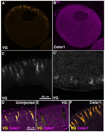
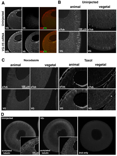
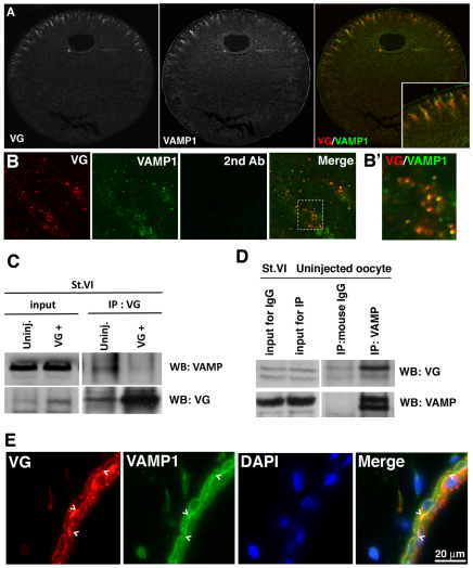
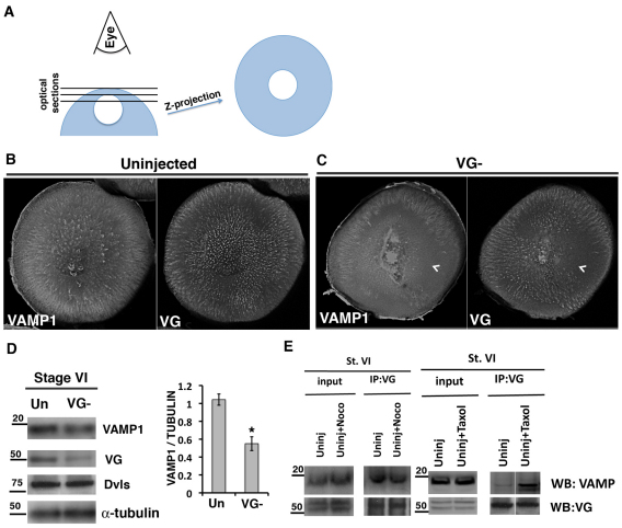
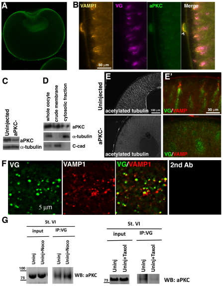
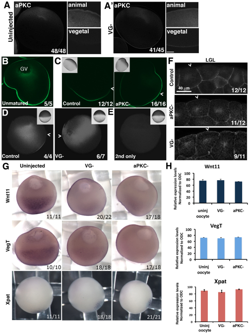
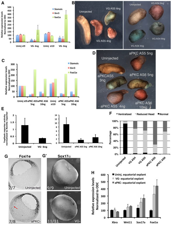
The Xenopus oocyte contains components of both the planar cell polarity and apical-basal polarity pathways, but their roles are not known. Here, we examine the distribution, interactions and functions of the maternal planar cell polarity core protein Vangl2 and the apical-basal complex component aPKC. We show that Vangl2 is distributed in animally enriched islands in the subcortical cytoplasm in full-grown oocytes, where it interacts with a post-Golgi v-SNARE protein, VAMP1, and acetylated microtubules. We find that Vangl2 is required for the stability of VAMP1 as well as for the maintenance of the stable microtubule architecture of the oocyte. We show that Vangl2 interacts with atypical PKC, and that both the acetylated microtubule cytoskeleton and the Vangl2-VAMP1 distribution are dependent on the presence of aPKC. We also demonstrate that aPKC and Vangl2 are required for the cell membrane asymmetry that is established during oocyte maturation, and for the asymmetrical distribution of maternal transcripts for the germ layer and dorsal/ventral determinants VegT and Wnt11. This study demonstrates the interaction and interdependence of Vangl2, VAMP1, aPKC and the stable microtubule cytoskeleton in the oocyte, shows that maternal Vangl2 and aPKC are required for specific oocyte asymmetries and vertebrate embryonic patterning, and points to the usefulness of the oocyte as a model to study the polarity problem.


Figures

Fig. 1.
Fig. 1.
Vangl2 and Celsr1 distribution in wild-type, full grown (stage 6) Xenopus oocytes. (A,B) Histological sections of stage 6 oocytes showing immunostaining pattern for Vangl2 (VG) protein (A) and Celsr1 protein (B). (C,C′), Higher magnification of animal (C) and vegetal (C') hemisphere distribution of Vangl2 protein. (D-F), Images of animal hemisphere sections co-immunostained for Vangl2 and Celsr1 in uninjected control sibling (D), Vangl2-depleted (VG–; E) and Celsr1-depleted (Celsr1–; F) stage 6 oocytes.
Fig. 2.
Fig. 2.
Vangl2 protein distributes with and maintains the acetylated microtubule cytoskeleton. (A) Co-immunostaining of sections from uninjected and Vangl2-V5 mRNA (20 pg)-injected stage 6 Xenopus oocytes using Vangl2 (VG) and V5 tag (V5) antibodies. (B) Co-immunostaining of sections of wild-type stage 6 oocytes for acetylated tubulin (aTub) and Vangl2 (VG) in the animal and vegetal hemispheres. (C) Co-immunostaining for acetylated tubulin and Vangl2 protein in sections of nocodazole-treated (5 μg/ml) and taxol-treated (2 μM/ml) stage 6 oocytes. (D) Optical sections (2.6 μm thickness) of stage 6 control and Vangl2-depleted (VG–) oocytes stained as whole mounts to show the acetylated tubulin cytoskeleton. Insets show higher magnification images of histological sections stained with the same antibody. Right-hand panel shows a control whole mount stained with secondary antibody only.
Fig. 3.
Fig. 3.
Vangl2 interacts with VAMP1. (A) Histological section of a stage 6 Xenopus oocyte co-immunostained for Vangl2 (VG) and VAMP1. (B,B′) Colocalization of Vangl2 (red) and VAMP1 (green) in the stage 6 oocyte cytoplasm. B′ shows a high magnification image (×100 oil objective with digital zoom) of the boxed area in B demonstrating the co-distribution of Vangl2 and VAMP1 (yellow). (C) Co-immunoprecipitation analysis of lysates from wild-type and Vangl2 mRNA (VG+; 500 pg)-injected stage 6 (St. VI) oocytes showing that endogenous Vangl2 protein (VG) interacts with endogenous VAMP1 protein (VAMP). Overexpressed Vangl2 reduces the total Vangl2 interaction with VAMP1. The expression level of VAMP1 in these samples is shown in the input lanes. (D) Co-immunoprecipitation analysis of an uninjected oocyte showing that endogenous VAMP1 protein interacts with endogenous Vangl2 protein. Mouse IgG serves as a negative control for non-specific binding of Xenopus oocyte lysates to the bead. (E) Co-immunostaining for Vangl2 (VG) and VAMP1 protein in histological sections of stage 45 tadpole skin. DAPI staining shows nuclei in the bilayered epidermis. Colocalization of VG and VAMP1 is most obvious on the basal side of the outer layer of the epithelium (chevrons, yellow staining in merge).
Fig. 4.
Fig. 4.
Vangl2 is required for VAMP1 protein levels. (A) Diagram showing the procedure for generating the z-stack images shown in B and C. (B,C) z-stacks of the entire animal hemisphere projected as a single image for control (B, Uninjected) and Vangl2-depleted (C, VG–) stage 6 Xenopus oocytes, co-immunostained for VAMP1 and Vangl2 (VG) proteins. Chevrons point to areas that are devoid of both VAMP1 and Vangl2. (D) Western blot of lysates from stage 6 control (Un) and Vangl2-depleted (VG–) oocytes probed for VAMP1 and Vangl2. Antibodies against alpha-tubulin and Xenopus Disheveleds (Dvls) are used as loading controls. Quantification is shown for VAMP1 versus alpha tubulin for this experiment. The experiment was repeated with a similar result (*P<0.02, determined by t-test). Error bars represent s.d. (E) Co-immunoprecipitation analyses showing that endogenous Vangl2 protein interacts with VAMP1 in control (Uninj) and nocodazole (Noco)-treated stage 6 (St. VI) oocyte lysates, and that the interaction is increased by taxol treatment.
Fig. 5.
Fig. 5.
aPKC regulates the distribution of the acetylated microtubule cytoskeleton and Vangl2-VAMP1 islands. (A) Histological section of a follicle-free (collagenase-treated) stage 6 Xenopus oocyte stained for aPKC protein. The nuclear staining is non-specific, as it is also observed in aPKC-depleted oocytes (not shown). (B) High magnification images of a histological section of the animal cortex of a control stage 6 oocyte co-immunostained for VAMP1, Vangl2 (VG) and aPKC. Chevron indicates a follicle cell. (C) Western blot showing aPKC protein levels in control and aPKC antisense oligonucleotide-injected (aPKC–) oocytes. (D) Western blot for aPKC using lysates from membrane and cytosolic fractions. Total α-tubulin is used as a marker for the cytosolic fraction and C-cadherin (C-cad) is used as a membrane fraction marker. (E,E′) Sections of control (upper panels) and aPKC-depleted (lower panels) stage 6 oocytes immunostained for acetylated tubulin (E) and Vangl2 (green) and VAMP1 (red) (E′). (F) Colocalization of Vangl2 (green) and VAMP1(red) proteins in aPKC-depleted oocyte cytoplasm. Right-hand panel shows incubation with secondary antibody only. (G) Co-immunoprecipitation analyses of lysates of control stage 6 (St. VI) oocytes showing that endogenous Vangl2 protein interacts with aPKC and that this interaction is not reduced by nocodazole (Noco) treatment (5 μg/ml) but is reduced by taxol treatment (2 μM/ml).
Fig. 6.
Fig. 6.
Vangl2 and aPKC are required for specific oocyte membrane and mRNA asymmetries. (A,A′) Low and high magnification images of hemisected whole-mount, progesterone-matured control (A) and Vangl2 depleted (VG–; A′) Xenopus oocytes stained for endogenous aPKC. (B,C) Localization of overexpressed GFP-LGL protein in anti-GFP immunostained histological sections of a stage 6 control oocyte (B) and progesterone-matured control and aPKC-depleted (PKC–) oocytes injected with GFP-LGL mRNA (C). Insets show bright-field images. Chevrons indicate the boundary between the animal and the vegetal hemispheres. (D) Comparison of the staining pattern of GFP-LGL in whole-mount anti-GFP immunostained control and Vangl2-depleted (VG–) progesterone-matured oocytes. Insets show the brightfield appearance of each oocyte and chevrons indicate the junction between animal and vegetal hemispheres. (E) Control for experiment shown in D showing a progesterone-matured oocyte that did not receive GFP-LGL mRNA injection. (F) GFP-LGL protein localization in histological sections of control, aPKC-depleted (aPKC–) and Vangl2-depleted (VG–) animal cells of embryos at the blastula stage, injected with GFP-LGL mRNA at the 2-cell stage. Chevrons mark the apical membrane of superficial cells in the animal cap. (G) In situ hybridization of sibling control, Vangl2-depleted (VG–) and aPKC-depleted (aPKC–) stage 6 oocytes probed for Wnt11, VegT and Xpat mRNAs. (H) Relative expression levels of Wnt11, VegT and Xpat mRNAs in control (uninj oocyte), Vangl2-depleted (VG–) and aPKC-depleted (aPKC–) oocytes determined by real-time RT-PCR (mean±s.d.).
Fig. 7.
Fig. 7.
Maternal Vangl2 and aPKC depletion disrupts the pattern of expression of specific zygotic genes. (A) Relative expression levels of Siamois, Xnr3 and Foxi1e mRNAs in control (Uninj) and Vangl2-depleted (VG–) embryos at the late blastula (st9) and early gastrula stages (st10) determined by real-time RT-PCR (mean±s.d.). (B) Phenotype of embryos derived from sibling control (Uninjected) and Vangl2-depleted oocytes (VG AS5, 4 and 6 ng oligonucleotide injected; VG AS4, 4, 6 and 8 ng oligonucleotide injected) at the early tailbud stage. (C) Relative expression levels of Siamois, Xnr3 and Foxi1e in control (Uninj) and aPKC-depleted embryos (AS5, 5 ng; AS6, 10 ng) determined by real-time RT-PCR at the late blastula and early gastrula stages. The experiment was repeated with a similar result. (D) Phenotype of embryos derived from sibling control (Uninjected) and aPKC-depleted oocytes (AS5, 3, 4 and 5 ng; AS6, 5 and 10 ng) at the early tailbud stage. (E) TOPflash reporter activation after injection into two dorsal vegetal cells of 8-cell stage control embryos compared with sibling Vangl2-depleted (left-hand graph) and aPKC-depleted (right-hand graph) embryos frozen at the late blastula stage, showing the reduction in canonical Wnt signaling activity (mean±s.d.). (F) Percentage of embryos with different phenotypes derived from Vangl2-depleted (AS4 and AS5), aPKC-depleted (AS5 and AS6) and sibling control at the tailbud stage. (G) Histological section of whole-mount stained, in situ hybridization of sibling control (upper) and aPKC-depleted (lower) early gastrulae using a Foxi1e probe. Arrow indicates ectopic expression of Foxi1e in the vegetal mass. (G′) Whole-mount in situ hybridization of sibling control (upper) and Vangl2-depleted (lower) early gastrulae using a Sox17a probe. (H) Relative expression levels of Xbra, Wnt11, Sox17a and Foxi1e in equatorial zones of control (Uninj.), Vangl2-depleted (VG–) and aPKC-depleted (aPKC–) embryos dissected at the late blastula stage and assayed by real-time RT-PCR (mean±s.d.).

References

    1. Aleu J., Blasi J., Solsona C., Marsal J. (2002). Calcium-dependent acetylcholine release from Xenopus oocytes: simultaneous ionic currents and acetylcholine release recordings. Eur. J. Neurosci. 16, 1442-1448 - PubMed
    1. Birsoy B., Kofron M., Schaible K., Wylie C., Heasman J. (2006). Vg 1 is an essential signaling molecule in Xenopus development. Development 133, 15-20 - PubMed
    1. Breckenridge R. A., Mohun T. J., Amaya E. (2001). A role for BMP signalling in heart looping morphogenesis in Xenopus. Dev. Biol. 232, 191-203 - PubMed
    1. Cha S. W., Heasman J. (2010). Using oocytes for Wnt signaling assays: paracrine assays and Wnt-conditioned medium. Methods 51, 52-55 - PubMed
    1. Cha S. W., Tadjuidje E., Tao Q., Wylie C., Heasman J. (2008). Wnt5a and Wnt11 interact in a maternal Dkk1-regulated fashion to activate both canonical and non-canonical signaling in Xenopus axis formation. Development 135, 3719-3729 - PubMed

Publication types

MeSH terms