. 2011 Oct;85(19):9877-86.
doi: 10.1128/JVI.05558-11. Epub 2011 Aug 3.

A Kaposi's sarcoma-associated herpesvirus-encoded ortholog of microRNA miR-155 induces human splenic B-cell expansion in NOD/LtSz-scid IL2Rγnull mice

Affiliations

A Kaposi's sarcoma-associated herpesvirus-encoded ortholog of microRNA miR-155 induces human splenic B-cell expansion in NOD/LtSz-scid IL2Rγnull mice

Isaac W Boss et al. J Virol. 2011 Oct.

Abstract

MicroRNAs (miRNAs) are small noncoding RNA molecules that function as posttranscriptional regulators of gene expression. Kaposi's sarcoma (KS)-associated herpesvirus (KSHV), a B-cell-tropic virus associated with KS and B-cell lymphomas, encodes 12 miRNA genes that are highly expressed in these tumor cells. One viral miRNA, miR-K12-11, shares 100% seed sequence homology with hsa-miR-155, an oncogenic human miRNA that functions as a key regulator of hematopoiesis and B-cell differentiation. So far, in vitro studies have shown that both miRNAs can regulate a common set of cellular target genes, suggesting that miR-K12-11 may mimic miR-155 function. To comparatively study miR-K12-11 and miR-155 function in vivo, we used a foamy virus vector to express the miRNAs in human hematopoietic progenitors and performed immune reconstitutions in NOD/LtSz-scid IL2Rγ(null) mice. We found that ectopic expression of miR-K12-11 or miR-155 leads to a significant expansion of the CD19(+) B-cell population in the spleen. Subsequent quantitative PCR analyses of these splenic B cells revealed that C/EBPβ, a transcriptional regulator of interleukin-6 that is linked to B-cell lymphoproliferative disorders, is downregulated when either miR-K12-11 or miR-155 is ectopically expressed. In addition, inhibition of miR-K12-11 function using antagomirs in KSHV-infected human primary effusion lymphoma B cells resulted in derepression of C/EBPβ transcript levels. This in vivo study validates miR-K12-11 as a functional ortholog of miR-155 in the context of hematopoiesis and suggests a novel mechanism by which KSHV miR-K12-11 induces splenic B-cell expansion and potentially KSHV-associated lymphomagenesis by targeting C/EBPβ.


Figures

Fig. 1.
Fig. 1.
The foamy virus vectors expressing miR-K12-11 or miR-155 and the empty-vector control used in this study. (A) Foamy virus vectors were constructed by inserting the pri-miRNA sequence downstream of a GFP cassette and a cytomegalovirus (CMV) promoter. LTR, long terminal repeat. (B) Schematic of miRNA sensor vectors containing two perfectly complementary binding sites. (C) miRNA expression and sensor vectors were cotransfected into 293T cells, and luciferase activity was measured at 72 h posttransfection. Results show that both miR-K12-11 and miR-155 expression vectors repressed luciferase activity >2-fold compared to no repression by the control.
Fig. 2.
Fig. 2.
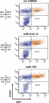
Engraftment of transduced CB CD34+ cells. Cells harvested from BM were analyzed by FACS using a human CD45-specific antibody. Human CD45+ cells were detected in all mice reconstituted with CD34+ human CB progenitors expressing either miR-K12-11, miR-155, or an empty control vector. A large percentage of the CD45+ cells also expressed GFP, as shown in the upper right quadrant of each histogram. Shown are representative dot plots for one animal from each group.
Fig. 3.
Fig. 3.
Ectopic miR-K12-11 and miR-155 expression in engrafted mice. (A) Ectopic miR-K12-11 was detected only in the miR-K12-11-engrafted animals in both BM and the spleen. (B) Ectopic miR-155 was detected above endogenous levels in miR-155-engrafted animals in both BM and the spleen. (C) The absolute copy numbers of miR-K12-11 in GFP-positive CD19+ splenocyte populations from engrafted mice are comparable to or lower than endogenous miR-K12-11 expression in the PEL cell line BCBL1, and therefore it is not overexpressed.
Fig. 4.
Fig. 4.
Cell lineage differentiation of human progenitors was not significantly altered by miRNA expression in BM. Cells harvested from the BM of mice expressing the empty vector (n = 4), miR-K12-11 (n = 7), or miR-155 (n = 8) were stained with antibodies specific for human (A) CD45+ leukocytes, (B) CD19+ B cells, (C) CD33+ monocytes, and (D) CD3+ T cells and analyzed by FACS. Each dot represents FACS analysis of one animal from each group, and the mean score of each group is shown as a solid horizontal line.
Fig. 5.
Fig. 5.
Ectopic expression of miR-K12-11 or miR-155 in human leukocytes during hematopoiesis leads to increased CD19+ B-cell expansion in the spleen. Splenocytes harvested from mice expressing the empty vector (n = 4), miR-K12-11 (n = 5), or miR-155 (n = 7) were stained with antibodies specific for human CD45+ leukocytes, CD19+ B cells, CD3+ T cells, and CD33+ monocytes and analyzed by FACS. (A) The fraction of human CD45+ leukocytes and CD19+ B cells was significantly higher (*, P < 0.05) in mice expressing either miR-K12-11 or miR-155 than in those expressing the empty control vector. No change was detected in the CD33+ monocyte or CD3+ T-cell population when either miRNA was expressed. Each dot represents FACS analysis of one animal from each group, and the mean score of each group is shown as a solid horizontal line. A P value (*) of 0.05 or less after a Student two-tailed t test was considered statistically significant. (B) Representative dot plots for flow cytometry analysis of splenocytes using hCD45+ and hCD19+ antibodies.
Fig. 6.
Fig. 6.
GFP-positive (miRNA-expressing) cells accounted for the overall increase in human CD45+ leukocytes and CD19+ B cells. Splenocytes harvested from mice expressing the empty vector (no-miRNA control, n = 4), miR-K12-11 (n = 5), or miR-155 (n = 7) were stained with antibodies specific for human CD45+ leukocytes or CD19+ B cells and analyzed for GFP expression by FACS. (A) The fraction of GFP-positive CD45+ human leukocytes was significantly higher (*, P < 0.05) in mice expressing either miR-K12-11 or miR-155 than in those expressing the empty control vector. (B) The fraction of GFP-positive CD19+ human B cells was significantly higher (*, P < 0.05) in mice expressing either miR-K12-11 or miR-155 than in those expressing the empty control vector. Each dot represents FACS analysis of one animal from each group, and the mean score of each group is shown as a solid horizontal line. A P value (*) of 0.05 or less after a Student two-tailed t test was considered statistically significant.
Fig. 7.
Fig. 7.
Immunohistochemical analysis of spleens revealed an increase in human CD19+ B-cell infiltrates in the splenic red pulp of mice expressing miR-K12-11 or miR-155. For IHC analysis, spleens were fixed, sectioned, and stained with a monoclonal antibody against human CD19. Photomicrographs of splenic sections at a ×40 magnification are shown at the top. The splenic red pulp regions are further magnified (×200) in the bottom panels to show the increased hCD19+ B-cell infiltrates (red staining) in the miRNA-expressing animals versus those in the no-miRNA control. Shown are representative sections from one animal in each group.
Fig. 8.
Fig. 8.
C/EBPβ is targeted by both miR-K12-11 and miR-155 in splenocytes and is regulated by miR-K12-11 in PEL. (A) The C/EBPβ 3′ UTR contains 1 seed match site for miR-K12-11 and miR-155. CDS, coding sequences. (B) The full-length C/EBPβ 3′ UTR was cloned (nucleotides 1233 to 1836) downstream of luciferase (pGL3-C/EBPβ) and cotransfected into 293T cells with increasing amounts (400 ng and 800 ng) of miR-155 or miR-K12-11 expression plasmids and a Renilla luciferase control vector. Transfection was normalized to Renilla luciferase values, and firefly values were graphed as relative light units. (C) RNA harvested from splenocytes from two separate animals from each group (empty-vector control, miR-K12-11, or miR-155) was analyzed by qRT-PCR for expression of C/EBPβ mRNA and normalized to GAPDH. (D) miR-K12-11 function in PEL cell lines BCBL1 and BC1 was inhibited using 25 nM 2′OMe antagomir specific for miR-K12-11. RNA was harvested from these cells, and derepression of C/EBPβ mRNA was analyzed by qPCR and normalized to GAPDH. Mock-transfected cells were used as a control. All experiments represent the average of three independent replicates and were repeated at least two times.

References

    1. Asou H., et al. 1998. Mechanisms of growth control of Kaposi's sarcoma-associated herpes virus-associated primary effusion lymphoma cells. Blood 91:2475–2481 - PubMed
    1. Bartel D. P. 2009. MicroRNAs: target recognition and regulatory functions. Cell 136:215–233 - PMC - PubMed
    1. Bolisetty M. T., Dy G., Tam W., Beemon K. L. 2009. Reticuloendotheliosis virus strain T induces miR-155, which targets JARID2 and promotes cell survival. J. Virol. 83:12009–12017 - PMC - PubMed
    1. Boss I. W., Plaisance K. B., Renne R. 2009. Role of virus-encoded microRNAs in herpesvirus biology. Trends Microbiol. 17:544–553 - PMC - PubMed
    1. Cai X., et al. 2005. Kaposi's sarcoma-associated herpesvirus expresses an array of viral microRNAs in latently infected cells. Proc. Natl. Acad. Sci. U. S. A. 102:5570–5575 - PMC - PubMed

Publication types