. 2011 Oct;85(19):9964-73.
doi: 10.1128/JVI.05291-11. Epub 2011 Aug 3.

Low-pH-dependent changes in the conformation and oligomeric state of the prefusion form of herpes simplex virus glycoprotein B are separable from fusion activity

Affiliations

Low-pH-dependent changes in the conformation and oligomeric state of the prefusion form of herpes simplex virus glycoprotein B are separable from fusion activity

Stephen J Dollery et al. J Virol. 2011 Oct.

Abstract

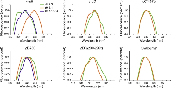
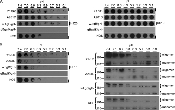
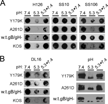
The cellular requirements for activation of herpesvirus fusion and entry remain poorly understood. Low pH triggers change in the antigenic reactivity of the prefusion form of the herpes simplex virus (HSV) fusion protein gB in virions, both in vitro and during viral entry via endocytosis (S. Dollery et al., J. Virol. 84:3759-3766, 2010). However, the mechanism and magnitude of gB conformational change are not clear. Here we show that the conformation and oligomeric state of gB with mutations in the bipartite fusion loops were similarly altered despite the fusion-inactivating mutations. Together with previous studies, this suggests that fusion loop mutants undergo conformational changes but are defective for fusion because they fail to make productive contact with the outer leaflet of the host target membrane. A direct, reversible effect of low pH on the structure of gB was detected by fluorescence spectroscopy. A soluble form of gB containing cytoplasmic tail sequences (s-gB) was triggered by mildly acidic pH to undergo changes in tryptophan fluorescence emission, hydrophobicity, antigenic conformation, and oligomeric structure and thus resembled the prefusion form of gB in the virion. In contrast, soluble gB730, for which the postfusion crystal structure is known, was only marginally affected by pH using these measures. The results underscore the importance of using a prefusion form of gB to assess the activation and extent of conformation change. Further, acidic pH had little to no effect on the conformation or hydrophobicity of gD or on gD's ability to bind nectin-1 or HVEM receptors. Our results support a model in which endosomal low pH serves as a cellular trigger of fusion by activating conformational changes in the fusion protein gB.


Figures

Fig. 1.
Fig. 1.
Effect of acid pH on tryptophan fluorescence of HSV glycoproteins. The indicated protein (10 to 20 μg/ml) was adjusted to a pH of 7.3 or 5.1. Indicated gB samples were acidified to pH 5.1 and then reneutralized to 7.4. Excitation was performed at 280 nm, and emission was recorded over 310 to 400 nm. Peak fluorescence is shown. Representative spectra from at least three independent experiments are shown.
Fig. 2.
Fig. 2.
Effect of low pH on the antigenic conformation of virion gD or soluble gD. (A) HSV-1 KOS virions were treated for 10 min at 37°C with medium buffered to pHs ranging from 7.4 to 5.2. Next, 105 PFU were blotted immediately to a nitrocellulose membrane. Blots were probed at neutral pH with gD-specific polyclonal antibody R7 or MAbs specific for gD (DL2, DL6, 1103, or DL11) or for gB (H1817 or H126), followed by horseradish peroxidase-conjugated goat secondary antibody. (B) Microtiter plates were coated with soluble gD(Δ290-299t) that had been treated with pH 7.4 or 4.7. MAb DL2, DL6, DL11, or 1103 to gD or control MAb to gC was added, followed by horseradish peroxidase-conjugated protein A and substrate. Values shown are the means of duplicate determinations.
Fig. 3.
Fig. 3.
Effect of low-pH pretreatment of gD on binding to HSV entry receptors HVEM or nectin-1. gD (Δ290-299t) was treated at pH 4.7, heated to 80°C, or left untreated. Increasing concentrations of gD were added to microtiter plates coated with HVEMt (A) or nectin-1t (B). Bound gD was detected with MAb DL6 followed by peroxidase-conjugated protein A and substrate. Absorbance was read at 410 nm. Each value represents the average of results for duplicate wells.
Fig. 4.
Fig. 4.
Effect of low pH on antigenic reactivity and hydrophobicity of different forms of soluble gB. (A) s-gB or gB730 was treated for 10 min at 37°C with medium buffered to pHs ranging from 7.4 to 5.1 and was blotted immediately to a nitrocellulose membrane. An equivalent amount of R69-reactive gB (50 to 100 μg) was blotted in each case. Blots were probed at neutral pH with gB-specific antibodies R69 (polyclonal) or H126, SS10, SS106, or DL16 (monoclonal), followed by horseradish peroxidase-conjugated goat secondary antibody. The exposures shown highlight the pH threshold of conformational change. (B) Proteins were added to 2% Triton X-114 that had been adjusted to the indicated pH. Samples were incubated for 10 min at 37°C and were centrifuged at 300 × g for 3 min. The aqueous supernatant phase or detergent phase was collected and diluted 20-fold in PBS. Samples were subjected to immunoprecipitation (H1817 for gB, DL6 for gD) followed by SDS-PAGE and immunoblotting (H1359 for gB, DL6 for gD).
Fig. 5.
Fig. 5.
Effect of low pH on the antigenic and oligomeric conformation of mutant gBs that are defective for fusion. (A) The indicated infected cell lysates were diluted in medium buffered to pHs ranging from 7.4 to 5.1. Samples were incubated at 37°C for 5 min and then blotted directly to nitrocellulose. Amounts of cell-associated virus that reacted equally to MAbs prior to pH treatment were blotted. Membranes were blocked and then incubated at neutral pH with anti-gB MAbs H126 (domain I) or SS10 (domain III) or (B, left panel) DL16 (trimer-specific). Similar results were obtained in at least three independent experiments per antibody. (B, right panel) Assay for sensitivity of oligomeric gB to detergent. The indicated infected cell lysates were treated with medium adjusted to pHs ranging from 7.4 to 5.0. Samples were adjusted to 1% SDS and were added to PAGE sample buffer containing 0.2% SDS and no reducing agent (“native” conditions). Unheated samples were resolved by PAGE. After transfer to nitrocellulose, membranes were blocked and incubated with gB-specific rabbit polyclonal antibody (R69). After incubation with horseradish peroxidase-conjugated goat-anti-rabbit antibody, enhanced chemiluminescent substrate was added and membranes were exposed to X-ray film. Molecular size markers in kilodaltons are indicated at the left. Positions of gB oligomer and monomer are indicated at the right. Each exposure (A and B) highlights the pH threshold of conformational change.
Fig. 6.
Fig. 6.
Reversibility of conformational changes in fusion loop mutants of gB. (A) The indicated infected cell lysates were treated with medium buffered to pH 7.4 or 5.3. For the indicated samples, the pH of 5.3 was neutralized to 7.4 for 10 min at 37°C and then the sample was blotted immediately to nitrocellulose. Membranes were probed at neutral pH with MAbs H126, SS10, SS106, or (B, left panel) DL16 followed by horseradish peroxidase-conjugated secondary antibody. Similar results were obtained in at least three independent experiments per antibody. (B, right panel) Cell-associated gB preparations were treated with the indicated pH conditions, treated with 1% SDS, and then analyzed by PAGE and immunoblotting with gB-specific polyclonal antibody R69. Images show gB of >181 kDa. Exposures shown highlight the reversibility of conformational change.

Similar articles

Cited by

References

    1. Arii J., et al. 2010. Non-muscle myosin IIA is a functional entry receptor for herpes simplex virus-1. Nature 467:859–862 - PubMed
    1. Arii J., et al. 2009. Entry of herpes simplex virus 1 and other alphaherpesviruses via the paired immunoglobulin-like type 2 receptor alpha. J. Virol. 83:4520–4527 - PMC - PubMed
    1. Atanasiu D., Saw W. T., Cohen G. H., Eisenberg R. J. 2010. Cascade of events governing cell-cell fusion induced by herpes simplex virus glycoproteins gD, gH/gL, and gB. J. Virol. 84:12292–12299 - PMC - PubMed
    1. Atanasiu D., et al. 2010. Bimolecular complementation defines functional regions of herpes simplex virus gB that are involved with gH/gL as a necessary step leading to cell fusion. J. Virol. 84:3825–3834 - PMC - PubMed
    1. Avitabile E., Forghieri C., Campadelli-Fiume G. 2009. Cross talk among the glycoproteins involved in herpes simplex virus entry and fusion: the interaction between gB and gH/gL does not necessarily require gD. J. Virol. 83:10752–10760 - PMC - PubMed

Publication types

MeSH terms