. 2011 Oct;85(20):10440-50.
doi: 10.1128/JVI.05408-11. Epub 2011 Aug 3.

Vesicular stomatitis virus expressing tumor suppressor p53 is a highly attenuated, potent oncolytic agent

Affiliations

Vesicular stomatitis virus expressing tumor suppressor p53 is a highly attenuated, potent oncolytic agent

Joshua F Heiber et al. J Virol. 2011 Oct.

Abstract

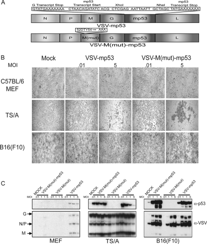
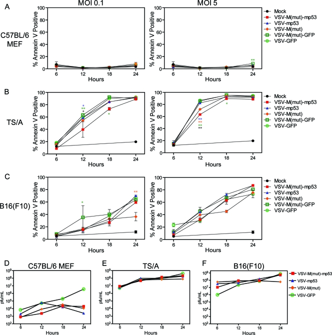
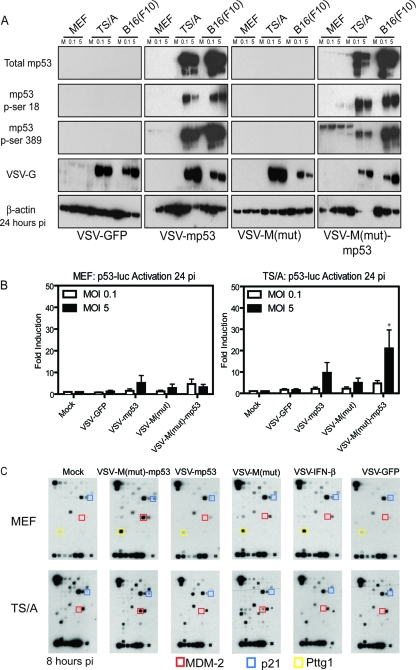
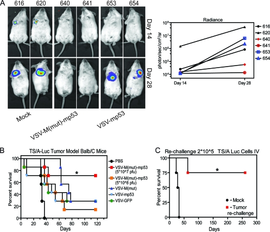
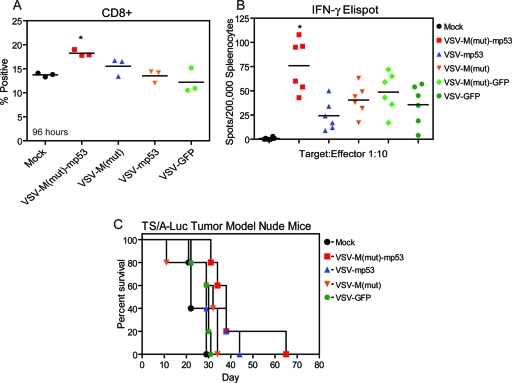
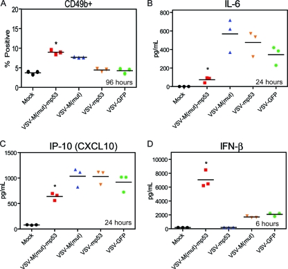
Vesicular stomatitis virus (VSV), a negative-strand RNA rhabdovirus, preferentially replicates in and eradicates transformed versus nontransformed cells and is thus being considered for use as a potential anticancer treatment. The genetic malleability of VSV also affords an opportunity to develop more potent agents that exhibit increased therapeutic activity. The tumor suppressor p53 has been shown to exert potent antitumor properties, which may in part involve stimulating host innate immune responses to malignancies. To evaluate whether VSV expressing p53 exhibited enhanced oncolytic action, the murine p53 (mp53) gene was incorporated into recombinant VSVs with or without a functional viral M gene-encoded protein that could either block (VSV-mp53) or enable [VSV-M(mut)-mp53] host mRNA export following infection of susceptible cells. Our results indicated that VSV-mp53 and VSV-M(mut)-mp53 expressed high levels of functional p53 and retained the ability to lyse transformed versus normal cells. In addition, we observed that VSV-ΔM-mp53 was extremely attenuated in vivo due to p53 activating innate immune genes, such as type I interferon (IFN). Significantly, immunocompetent animals with metastatic mammary adenocarcinoma exhibited increased survival following treatment with a single inoculation of VSV-ΔM-mp53, the mechanisms of which involved enhanced CD49b+ NK and tumor-specific CD8+ T cell responses. Our data indicate that VSV incorporating p53 could provide a safe, effective strategy for the design of VSV oncolytic therapeutics and VSV-based vaccines.


Figures

Fig. 1.
Fig. 1.
VSV-M(mut)-mp53 and VSV-mp53 retain oncolytic ability and express mp53. (A) Schematic representation of VSV p53 constructs. (B) Bright-field microscopy of VSV-M(mut)-mp53- and VSV-mp53-infected cells at MOIs of 0.01 and 5 24 h postinfection. Mock, mock infection. (C) Immunoblot analysis for VSV and mp53 protein expression in infected C57BL/6 MEFs and TS/A or B16(F10) cells infected at MOIs of 0.01, 0.1, 1, and 5 24 h postinfection.
Fig. 2.
Fig. 2.
VSV-M(mut)-mp53 and VSV-mp53 replication in vitro. C57BL/6 MEFs and TS/A or B16(F10) cells were infected with rVSVs at an MOI of 0.1, 1, or 5. Cells and supernatants were collected 6, 12, 18, and 24 h postinfection. (A, B, and C) Cell death at each time point as determined by annexin V-propidium iodide staining. (D, E, and F) Supernatants were analyzed by the standard plaque assay to determine the replication of rVSVs. (Two-way analysis of variance [ANOVA]/Bonferroni posttest: *, P < 0.001; **, P < 0.0001; blue asterisks, VSV-M(mut)-mp53 versus VSV-mp53; orange asterisks, VSV-M(mut)-mp53 versus VSV-ΔM; green asterisks, VSV-M(mut)-mp53 versus VSV-M(mut)-GFP; black asterisks, VSV-M(mut)-mp53 versus VSV-GFP). The error bars indicate standard deviations.
Fig. 3.
Fig. 3.
Expressed mp53 is functional. (A) C57BL/6 MEFs and TS/A or B16(F10) cells were infected with rVSV at MOIs of 0.1 and 5. The cells were lysed 24 h postinfection (pi) and used for immunoblot analysis. (B) C57BL/6 MEFs or TS/A cells were transfected with a p53 luciferase reporter and then infected at an MOI of 0.1 or 5. Luciferase levels were assessed 24 h postinfection. (*, P = 0.0366; one-way ANOVA.) The error bars indicate standard deviations. (C) C57BL/6 MEFs or TS/A cells were infected at an MOI of 10 or 1, respectively, and mp53 target gene mRNA expression was determined.
Fig. 4.
Fig. 4.
VSV-M(mut)-mp53 protects immunocompetent mice from TS/A-luc formation. (A) Mock- or rVSV-treated mice were anesthetized and injected intraperitoneally (i.p.) with a luciferin substrate, and luciferase activity was detected using the IVIS. Representative images from mock-, VSV-M(mut)-mp53-, and VSV-mp53-treated mice are shown. Luciferase radiance is an indicator of tumor burden and is quantified using Living Image software (right). (B) BALB/c mice (n = 7) were initially given 1 × 10⁵ TS/A-luc cells i.v. on day 0 and were then either mock or rVSV treated [5 × 10⁷ PFU or 5 × 10⁸ PFU VSV-M(mut)-mp53 only] on day 3. The mice were then monitored for survival (*, P = 0.0045; log rank test). (C) Previously treated or naïve mice (n = 4) were challenged with 2 × 10⁵ TS/A-luc cells on day 120 (day 0 for rechallenge), and survival was monitored (*, P = 0.0091; log rank test).
Fig. 5.
Fig. 5.
VSV-M(mut)-mp53 modulates T cells and is attenuated in athymic BALB/c nude mice. (A) BALB/c mice (n = 3) were infected with 5 × 10⁷ PFU rVSV; 96 h postinfection, the spleens were removed and total splenocytes were assessed for the percentage of CD8+ cells by flow cytometry (*, P = 0.0069; one-way ANOVA). (B) BALB/c mice (n = 6) were initially given 1 × 10⁵ TS/A-luc cells i.v. on day 0 and were then either mock treated or treated with 5 × 10⁷ PFU rVSV on day 3. On day 13, the mice were sacrificed, and total splenocytes were used for an IFN-γ ELISPOT with mitomycin C-treated TS/A-luc cells as the target (*, P < 0.0001; one-way ANOVA). Horizontal lines indicate means of results. (C) Athymic BALB/c nude mice (n = 5) were injected with 1 × 10⁵ TS/A-luc cells on day 0, mock treated or treated with 5 × 10⁷ PFU rVSV on day 3, and then observed for survival (P = 0.2077; log rank test).
Fig. 6.
Fig. 6.
VSV-M(mut)-mp53 modulates NK cell and cytokine profiles. (A, B, and C) BALB/c mice (n = 6) were treated with 5 × 10⁷ PFU rVSV i.v.; 24 and 96 h postinfection, splenocytes and sera were collected. (A) The splenocytes were stained for flow cytometry to determine the number that were CD49b+ (*, P < 0.0001; one-way ANOVA). (B and C) Sera were used for a multiplex ELISA in which IL-6 (B) and IP-10 (C) showed significant differences (*, P < 0.0001; one-way ANOVA). (D) BALB/c mice (n = 3) were infected with 5 × 10⁷ PFU rVSV. Six hours postinfection, sera were collected, and ELISA was used to assess IFN-β (*, P = 0.0003; one-way ANOVA). Horizontal lines indicate means of results.

Similar articles

Cited by

References

    1. Adkins B., Bu Y., Cepero E., Perez R. 2000. Exclusive Th2 primary effector function in spleens but mixed Th1/Th2 function in lymph nodes of murine neonates. J. Immunol. 164:2347–2353 - PubMed
    1. Angelo L. S., Talpaz M., Kurzrock R. 2002. Autocrine interleukin-6 production in renal cell carcinoma: evidence for the involvement of p53. Cancer Res. 62:932–940 - PubMed
    1. Angiolillo A. L., et al. 1995. Human interferon-inducible protein 10 is a potent inhibitor of angiogenesis in vivo. J. Exp. Med. 182:155–162 - PMC - PubMed
    1. Arai K., Liu Z. X., Lane T., Dennert G. 2002. IP-10 and Mig facilitate accumulation of T cells in the virus-infected liver. Cell Immunol. 219:48–56 - PubMed
    1. Balachandran S., Barber G. N. 2004. Defective translational control facilitates vesicular stomatitis virus oncolysis. Cancer Cell 5:51–65 - PubMed

Publication types

MeSH terms

Substances