. 2011 Oct;85(19):10021-30.
doi: 10.1128/JVI.05107-11. Epub 2011 Aug 3.

Crystal structures of enterovirus 71 3C protease complexed with rupintrivir reveal the roles of catalytically important residues

Affiliations

Crystal structures of enterovirus 71 3C protease complexed with rupintrivir reveal the roles of catalytically important residues

Jing Wang et al. J Virol. 2011 Oct.

Abstract

EV71 is the primary pathogenic cause of hand-foot-mouth disease (HFMD), but an effective antiviral drug currently is unavailable. Rupintrivir, an inhibitor against human rhinovirus (HRV), has potent antiviral activities against EV71. We determined the high-resolution crystal structures of the EV71 3C(pro)/rupintrivir complex, showing that although rupintrivir interacts with EV71 3C(pro) similarly to HRV 3C(pro), the C terminus of the inhibitor cannot accommodate the leaving-group pockets of EV71 3C(pro). Our structures reveal that EV71 3C(pro) possesses a surface-recessive S2' pocket that is not present in HRV 3C(pro) that contributes to the additional substrate binding affinity. Combined with mutagenic studies, we demonstrated that catalytic Glu71 is irreplaceable for maintaining the overall architecture of the active site and, most importantly, the productive conformation of catalytic His40. We discovered the role of a previously uncharacterized residue, Arg39 of EV71 3C(pro), that can neutralize the negative charge of Glu71, which may subsequently assist deprotonation of His40 during proteolysis.


Figures
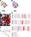
Fig. 1.
Fig. 1.
Overall structure of EV71 3Cpro/rupintrivir complex. (A) Ribbon model of unliganded EV71 3Cpro with annotated secondary structures. The β-ribbon adopts the open conformation. (B) Ribbon model of rupintrivir (carbon is in orange) binding to EV71 3Cpro with annotated secondary structures. The β-ribbon adopts the closed conformation. (C) Rupintrivir bound to EV71 3Cpro. The active site of the protease is rendered as a semitransparent solvent-accessible surface color coded at residues (involving substrate binding) according to amino acid conservation among the picornaviral 3Cpro of known structure. Residues indicated in red are invariant. Increasing amino acid variation at residues is indicated by progressively fading red to white, with white indicating the least conserved residues. The carbons of the bound rupintrivir are colored in white; the ordered waters are red spheres. (D) Structure-based multiple-sequence alignment of 3C proteases from different picornaviruses. The secondary structure is shown at the top. Invariant residues are white with a red background; conserved residues are shown in red font. The residues involved in the interactions with rupintrivir are marked below the sequences. Residues contacting with P1′, P1, P2, P3, and P4 of rupintrivir are blue, red, purple, brown, and green bars, respectively. (E) Structural formula of rupintrivir color coded as described for panel D. Residues involving the binding with rupintrivir are listed and color coded.
Fig. 2.
Fig. 2.
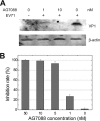
Inhibition of EV71 by rupintrivir (AG7088). The efficacy of rupintrivir against EV71 was evaluated in antiviral assays. The data showed that rupintrivir was effective against EV71 (isolate BJ/CHN/2008) with a mean EC50 of ∼1 nM, which is comparable to the antiviral activities of rupintrivir against HRV or other picornaviruses. No obvious cytotoxicity was found by adding rupintrivir to RD cells. The antiviral activity of rupintrivir was further demonstrated using Western blotting and RT-PCR analyses. RD cells infected with EV71 showed less quantity of VP1 protein than that detected in the presence rupintrivir (1 or 10 nM). RT-PCR experiments detecting the quantities of viral RNAs in RD cells show that rupintrivir suppresses EV71 replication in a dose-dependent manner, which is consistent with the results from antiviral assays. (A) Cell lysates (40 μg protein per lane) were prepared from either mock-infected (lane 1) or EV71-infected RD cells at 24 h postinfection and resolved with 12% SDS-PAGE. Western blot analysis for VP1 or β-actin was conducted. Lane 1, mock-infected cells not treated with rupintrivir (AG7088); lanes 2 to 4, cells were treated with 1, 10, or 0 nM AG7088, respectively. The bands corresponding to EV71 VP1 are indicated. The expression of β-actin was used to control equal protein loading. (B) Cells seeded in 24-well plates were treated with different concentrations of AG7088 and infected with EV71 at an MOI of 0.1. Twenty-four h postinfection viral RNAs were extracted from cells, and real-time RT-PCR was performed. The inhibition rate (percent inhibition of viral RNA) is plotted as a function of inhibitor concentrations. The plotted data are averages from triplicates.
Fig. 3.
Fig. 3.
Interactions between EV71 3Cpro and rupintrivir. (A) Superimposition of the stick models of EV71 3Cpro (carbon in orange), HRV 3Cpro (carbon in gray; PDB no. 1cqq), and CVB 3Cpro(carbon in teal; PDB no. 2zu3) bound by different inhibitors. The superimposed oxyanion holes are indicated with an arrow. (B) Stick model of EV71 3Cpro/rupintrivir (carbon in orange) superimposed with the stick model of HRV 3Cpro/rupintrivir (carbon in gray; PDB no. 1cqq). Residues on the differently orientated tight loops (aa 105 to 111) are labeled. The dashed lines indicate the electrostatic interaction between Asn107 and the C-terminal carbonyl oxygen of rupintrivir in HRV 3Cpro. (C) Stick model of EV71 3Cpro/rupintrivir (carbon in orange) and the superimposed stick model of HRV 3Cpro/rupintrivir (carbon in gray). The 4-fluoroPhe side chain of P2 of rupintrivir is buried more shallowly in the S2 pocket in EV71 3Cpro than in HRV 3Cpro. The basic residues Arg39 and Lys130 form the closed distal end of the S2 pocket in EV71 3Cpro and interact with the side chain fluoride of the P2 residue of rupintrivir. (D) Electrostatic surface of proteases active sites bound by inhibitors with annotated substrate specificity pockets. The position of the shallow pit defined by a loop of aa 105 to 111 is indicated with a black arrow. Image 1 shows that the distal end of the S2 specificity pocket is closed in EV71 3Cpro, whereas the distal end of the S2 pocket is open in other picornaviral 3C proteases; image 2, rupintrivir bound to EV71 3Cpro; image 3, rupintrivir bound to HRV 3Cpro (PDB no. 1cqq); image 4, inhibitor TG-0204998 bound to CVB 3Cpro (PDB no. 2zu3). The peptide substrate of FMDV 3Cpro (PDB no. 2wv4) is modeled in the substrate binding cleft in EV71 3Cpro by superimposition, showing that the shallow pit next to the S1′ pocket can serve as the S2′ pocket accommodating the hydrophobic P2′ side chain.
Fig. 4.
Fig. 4.
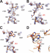
Structure of the active sites. (A, B, and C) Final 2Fo-Fc electron densities (1.5-s contour) at the active site and the superimposed final model of EV71 3Cpro mutants H133G, E71A, and E71D. The hydrogen bond network of the active site is indicated with dashed lines, and each hydrogen bond is numbered. Residues involved in the hydrogen bond network of the active site are annotated. Mutated residues are indicated in red. (D) Stick model of the EV71 3Cpro mutant E71D active site (carbon in orange) and the superimposed FMDV 3Cpro active site (carbon in gray). Catalytically important residues are indicated. Distances between catalytic His and Asp are indicated with dashed lines.
Fig. 5.
Fig. 5.
Mutagenic studies of residues Arg39 and Asn69. Specificity constants (kcat/Km) of WT EV71 3Cpro and mutants R39K, R39E, R39T, N69S, and N69D are shown. Error bars represent standard errors from the nonlinear regression analysis. The data show that besides the canonical catalytic triad, the residues Asn69 and Arg39 also are important to proteolysis.
Fig. 6.
Fig. 6.
Proteolysis mechanism of EV71 3Cpro involving the neutralizer Arg39. The catalytically important residues are black. The substrate is green. The electron relays are indicated by red arrows. Thicker dashed lines indicate stronger interactions, and thinner dashed lines indicate weaker interactions.

Similar articles

Cited by

References

    1. Adams P. D., et al. 2010. PHENIX: a comprehensive Python-based system for macromolecular structure solution. Acta Crystallogr. D Biol. Crystallogr 66:213–221 - PMC - PubMed
    1. Allaire M., Chernaia M. M., Malcolm B. A., James M. N. 1994. Picornaviral 3C cysteine proteinases have a fold similar to chymotrypsin-like serine proteinases. Nature 369:72–76 - PubMed
    1. Ash E. L., et al. 2000. Unusual 1H NMR chemical shifts support (His) C(epsilon) 1.O==C H-bond: proposal for reaction-driven ring flip mechanism in serine protease catalysis. Proc. Natl. Acad. Sci. U. S. A. 97:10371–10376 - PMC - PubMed
    1. Bergmann E. M., et al. 1999. Crystal structure of an inhibitor complex of the 3C proteinase from hepatitis A virus (HAV) and implications for the polyprotein processing in HAV. Virology 265:153–163 - PubMed
    1. Bergmann E. M., Mosimann S. C., Chernaia M. M., Malcolm B. A., James M. N. 1997. The refined crystal structure of the 3C gene product from hepatitis A virus: specific proteinase activity and RNA recognition. J. Virol. 71:2436–2448 - PMC - PubMed

Publication types

MeSH terms

LinkOut - more resources