. 2011 Aug 3;31(31):11100-9.
doi: 10.1523/JNEUROSCI.6752-10.2011.

Suppression of a neocortical potassium channel activity by intracellular amyloid-β and its rescue with Homer1a

Affiliations

Suppression of a neocortical potassium channel activity by intracellular amyloid-β and its rescue with Homer1a

Kenji Yamamoto et al. J Neurosci. .

Abstract

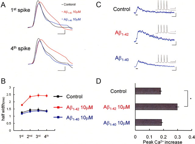
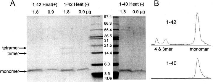
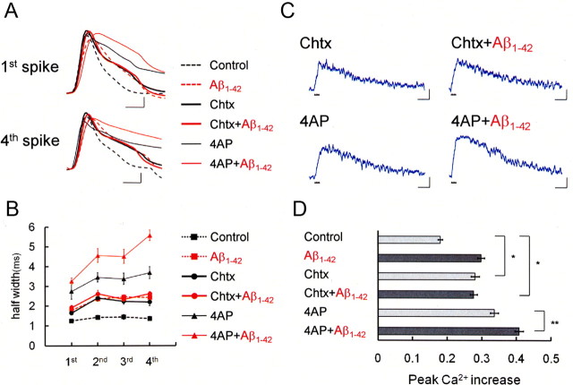
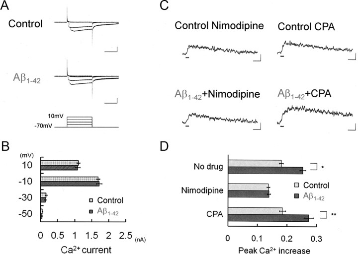
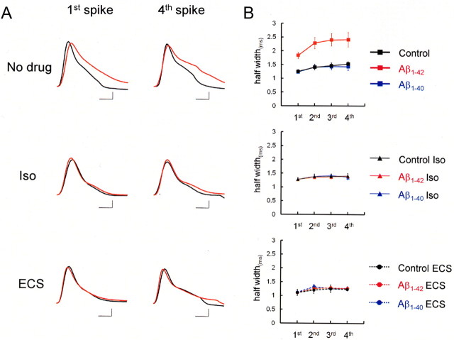
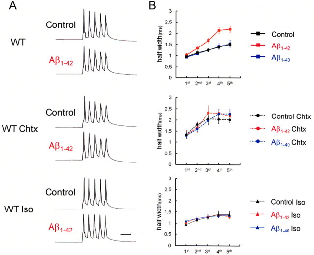
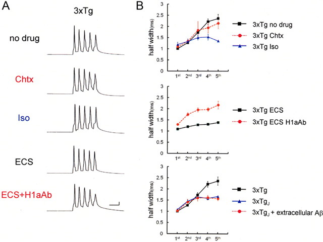
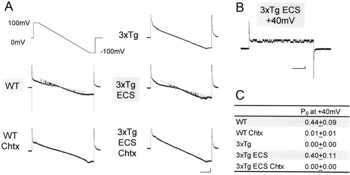
It is proposed that intracellular amyloid-β (Aβ), before extracellular plaque formation, triggers cognitive deficits in Alzheimer disease (AD). Here we report how intracellular Aβ affects neuronal properties. This was done by injecting Aβ protein into rat and mouse neocortical pyramidal cells through whole-cell patch pipettes and by using 3xTg AD model mice, in which intracellular Aβ is accumulated innately. In rats, intracellular application of a mixed Aβ(1-42) preparation containing both oligomers and monomers, but not a monomeric preparation of Aβ(1-40), broadened spike width and augmented Ca(2+) influx via voltage-dependent Ca(2+) channels in neocortical neurons. Both effects were mimicked and occluded by charybdotoxin, a blocker of large-conductance Ca(2+)-activated K(+) (BK) channels, and blocked by isopimaric acid, a BK channel opener. Surprisingly, augmented Ca(2+) influx was caused by elongated spike duration, but not attributable to direct Ca(2+) channel modulation by Aβ(1-42). The Aβ(1-42)-induced spike broadening was blocked by electroconvulsive shock (ECS), which we previously showed to facilitate BK channel opening via expression of the scaffold protein Homer1a. In young 3xTg and wild mice, we confirmed spike broadening by Aβ(1-42), which was again mimicked and occluded by charybdotoxin and blocked by ECS. In Homer1a knock-out mice, ECS failed to block the Aβ(1-42) effect. Single-channel recording on BK channels supported these results. These findings suggest that the suppression of BK channels by intracellular Aβ(1-42) is a possible key mechanism for early dysfunction in the AD brain, which may be counteracted by activity-dependent expression of Homer1a.


Figures

Figure 1.
Figure 1.
Intracellular infusion of Aβ1-42 broadens spike width and augmented Ca2+ influx in rat neocortical pyramidal neurons. A, Action potentials evoked in neurons injected with Aβ. The first and fourth action potentials in spike trains are shown. Recordings taken from the control neuron (black), an Aβ1-42-injected neuron (red), and an Aβ1-40-injected neuron (blue) are superimposed to clarify the spike broadening in Aβ1-42-injected neurons. Calibration: 1 ms, 20 mV. B, Average spike half-width in four-spike trains, showing that injection of Aβ1-42 (10 μm; red square; n = 6), but not that of Aβ1-40 (10 μm; blue square; n = 6), broadened spike width compared with control neurons (black square; n = 6). C, Ca2+ increases induced by four-spike trains (inset) in control neurons and in neurons injected with 10 μm1-42 or 10 μm1-40. Calibration: 500 ms, −0.1ΔF380/F360. Inset, Specimen recording of a spike train at 18 Hz that was used for the shown Ca2+ measurements. The timing of each spike is shown by a small black triangle below the trace. Note the difference in time scale. Calibration: 100 ms, 20 mV. D, Summary diagram demonstrating average Ca2+ increases. Injection of Aβ1-42 enhanced spike-induced Ca2+ increases. *p < 0.0001.
Figure 2.
Figure 2.
Detection of Aβ1-42 oligomers in the pipette solution. A, Aβ1-42 and Aβ1-40 were taken from the pipette solution and prepared for SDS-PAGE. Two different amounts (1.8 and 0.9 μg) were used. Samples were heated [Heat(+)] before loading for controls or left without heating [Heat(−)]. Based on the molecular-weight marker, the positions for monomer, trimer, and tetramer were determined. For Aβ1-42 only, in addition to the monomer, bands are also positive at the trimer and tetramer positions, albeit to much lesser extents than the monomer band [1–42 Heat(−), 1.8 μg]. B, Band density profiles. By applying the Plot Lanes function (ImageJ software) to scanned gel images, density profiles were drawn for the lanes loaded with unheated Aβ1-42 [1–42 Heat(−), 1.8 μg] and Aβ1-40 [1–40 Heat(−), 1.8 μg]. Relative optical density was plotted in an arbitrary unit for each lane. The top-to-bottom direction in the lane is reflected to the left-to-right direction in this diagram. Compared with the peak density for the monomer band, those for trimer and tetramer were much lower for Aβ1-42 and undetectable for Aβ1-40.
Figure 3.
Figure 3.
Intracellular Aβ1-42 enlarges spike width by suppressing BK channels, thereby increasing spike-induced Ca2+ entry. A, First and fourth action potentials during spike trains after application of the BK channel blocker Chtx (50 nm) or the A-type K+ channel blocker 4-AP (5 mm) in a control cell (Chtx: thick black trace, n = 7; 4-AP: thin black trace, n = 5) and an Aβ1-42-injected cell (Chtx+Aβ1-42: thick red trace, n = 4; 4-AP+Aβ1-42: thin red trace, n = 4). The action potentials in the control cell (black dashed trace) and Aβ1-42-injected cell (red dashed trace) shown in Figure 1 are superimposed. Calibration: 1 ms, 20mV. B, Averaged spike half-width during four-spike trains with no blocker (black square, Control; red square, Aβ1-42), with charybdotoxin (black circle, Control; red circle, Aβ1-42) and with 4-AP (black triangle, Control; red triangle, Aβ1-42). C, Spike-induced Ca2+ increase under application of charybdotoxin or 4-AP in control or Aβ1-42-injected neurons. Calibration: 500 ms, −0.1ΔF380/F360. D, Summary diagram demonstrating average Ca2+ increases in the control, charybdotoxin, and 4-AP groups, each with and without Aβ1-42. In B and D, charybdotoxin, but not 4-AP, mimicked and occluded the effect of Aβ1-42. *p < 0.0001; **p < 0.01.
Figure 4.
Figure 4.
Failure of intracellular Aβ1-42 to directly modulate Ca2+ channels. A, Ca2+ currents with 1 μm1-42 (n = 10) or without Aβ1-42 (Control; n = 7) infused. Calibration: 100 ms, 1 nA. B, The peak amplitude of Ca2+ current (elicited by voltage steps to −50, −30, −10, and 10 mV) was compared. Aβ1-42 infusion has no effects on Ca2+ currents. C, The voltage-dependent Ca2+ channel blocker nimodipine (20 μm; Control, n = 5; Aβ1-42, n = 5), but not the Ca2+ store depleter CPA (30 μm; Control, n = 6; Aβ1-42, n = 5), abolishes the enhancement of spike-induced Ca2+ increase by Aβ1-42. Ca2+ channel activation becomes stronger with Aβ1-42, rather indirectly, because of BK channel suppression by Aβ1-42 (Fig. 3). Calibration: 500 ms, −0.1ΔF380/F360. D, Peak amplitude of spike-induced Ca2+ increases in the presence of nimodipine or CPA, indicating that Ca2+ release channels are not involved at all. *p < 0.001; **p < 0.01.
Figure 5.
Figure 5.
ECS blocked Aβ1-42-mediated suppression of BK channels in rat neocortical neurons. A, First and fourth spike in spike trains under the application of the BK channel opener isopimaric acid (Iso; 10 μm) or after ECS, in a control neuron (black) and an Aβ1-42-injected neuron (red). The same spike broadening as observed with fura-2 was confirmed without fura-2 in patch pipettes (Fig. 1). Note differences to the same extent in spike width between cells with (red line) and without intracellular Aβ1-42 (black line, No drug). Both isopimaric acid and ECS prevented Aβ1-42 from broadening spike width. Calibration: 1 ms, 20mV. B, Averaged spike half-width during four-spike trains (black, Control; red, Aβ1-42; blue, Aβ1-40) with No drug (top), with Iso (middle), and after ECS (bottom). Each point is based on an average over four to eight trials.
Figure 6.
Figure 6.
1-42 injection into neurons in slices obtained from wild mice caused spike broadening. A, Recordings of spike trains induced by brief current injections at 100 Hz into neurons in slices obtained from wild mice. With Aβ1-42 injected intracellularly, spike half-widths in later spikes were significantly larger than in no-Aβ controls. Charybdotoxin mimics and occludes and Iso blocks the actions of Aβ1-42, indicating that spike broadening is caused by Aβ1-42-mediated blockade of BK channels. Calibration: 20 ms, 20 mV. B, Averaged spike half-width during the five-spike train (black, Control; red, Aβ1-42; blue, Aβ1-40) with no drug (top), with Chtx (middle), and with Iso (bottom). Each point is an average of four to six trials.
Figure 7.
Figure 7.
Blocking effects of ECS on Aβ1-42 was absent in H1aKO mice. A, ECS counteracts Aβ1-42-mediated spike broadening in wild-type mice (WT ECS) but not in H1aKO mice (H1aKO, H1aKO ECS). Calibration: 20 ms, 20 mV. B, Averaged spike half-width during spike trains (black, Control; red, Aβ1-42) with ECS in wild mice (top), without ECS in H1aKO mice (middle), and with ECS in H1aKO mice (bottom). Each point is an average of four to six trials.
Figure 8.
Figure 8.
Spike broadening in 3xTg neurons. A, Recordings of spike trains in the 3xTg mice at 4 months of age, exhibiting spike broadening. The top recording shows spike broadening in the naive condition (no drug, n = 6). The addition of charybdotoxin failed to broaden spikes further (n = 4), exhibiting occlusion of the blocker effect. Iso blocked the spike broadening (n = 4). The bottom two recordings show spike trains in neurons from 3xTg mice after ECS. ECS on 3xTg mice blocked the spike broadening, which was cancelled out by injection of anti-Homer1a antibody (0.4 μg/ml; ECS, n = 6 vs ECS +H1aAb, n = 7). Calibration: 20 ms, 20 mV. B, Averaged spike width in 3xTg cells. Top, Spike half-width is shown for naive 3xTg neurons (No drug; black square), those with BK channel blockade (Chtx; red circle), and those with Iso (blue triangle). Middle, Averaged spike half-width after ECS with H1aAb (ECS+H1aAb; red circle) or without (ECS; black square). Bottom, Spike half-width recorded in neurons from juvenile 3xTg (16–18 d old; 3xTgj), in which intracellular Aβ has not yet been accumulated. Spike width was smaller in 3xTgj neurons (3xTgj; blue triangle) than in 3xTg neurons (3xTg; black square) and remained at a range comparable with that in wild controls. Extracellular application of Aβ1-42 (1 μm) to 3xTgj cells failed to broaden spikes (3xTgj+extracellular Aβ; red circle).
Figure 9.
Figure 9.
Recovery of single BK current by ECS in 3xTg mice. A, A ramp-voltage command from +100 to −100 mV (top left) induces single BK currents in wild mice (WT; n = 6) and 3xTg mice after ECS (3xTg ECS; n = 5), but not in 3xTg mice without ECS (3xTg; n = 6). These single currents were blocked by Chtx (n = 5; 3xTg ECS Chtx, n = 4). The dotted lines indicate the open or closed states. Calibration: 200 ms, 10 pA. B, Single BK currents were confirmed by a rectangle voltage command from 0 to +40 mV in 3xTg mice after ECS. This voltage step was used, since the open probability hereby was found close to ∼50%. Calibration: 200 ms, 5 pA. C, Open probability (Po) based on recordings with step commands to +40 mV. Each point is an average of four to six trials.

Similar articles

Cited by

References

    1. Abramov E, Dolev I, Fogel H, Ciccotosto GD, Ruff E, Slutsky I. Amyloid-β as a positive endogenous regulator of release probability at hippocampal synapses. Nat Neurosci. 2009;12:1567–1576. - PubMed
    1. Ango F, Prézeau L, Muller T, Tu JC, Xiao B, Worley PF, Pin JP, Bockaert J, Fagni L. Agonist-independent activation of metabotropic glutamate receptors by the intracellular protein Homer. Nature. 2001;411:962–965. - PubMed
    1. Billings LM, Oddo S, Green KN, McGaugh JL, LaFerla FM. Intraneuronal Aβ causes the onset of early Alzheimer's disease-related cognitive deficits in transgenic mice. Neuron. 2005;45:675–688. - PubMed
    1. Brakeman PR, Lanahan AA, O'Brien R, Roche K, Barnes CA, Huganir RL, Worley PF. Homer: a protein that selectively binds metabotropic glutamate receptors. Nature. 1997;386:284–288. - PubMed
    1. Cirrito JR, Yamada KA, Finn MB, Sloviter RS, Bales KR, May PC, Schoepp DD, Paul SM, Mennerick S, Holtzman DM. Synaptic activity regulates interstitial fluid amyloid-β levels in vivo. Neuron. 2005;48:913–922. - PubMed

Publication types

MeSH terms

Substances

LinkOut - more resources