. 2011 Aug 3;31(31):11118-25.
doi: 10.1523/JNEUROSCI.1709-11.2011.

Two transcription factors can direct three photoreceptor outcomes from rod precursor cells in mouse retinal development

Affiliations

Two transcription factors can direct three photoreceptor outcomes from rod precursor cells in mouse retinal development

Lily Ng et al. J Neurosci. .

Abstract

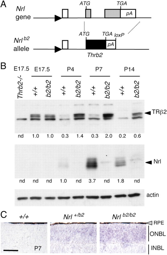
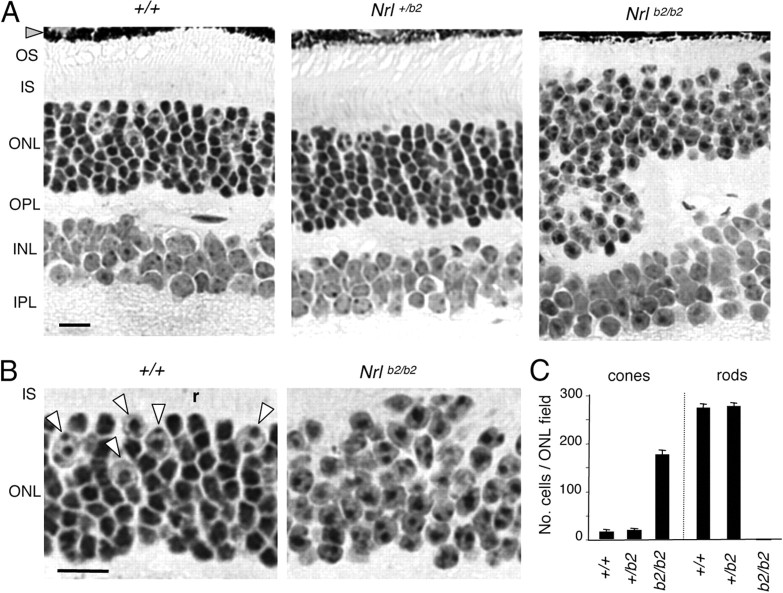
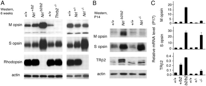
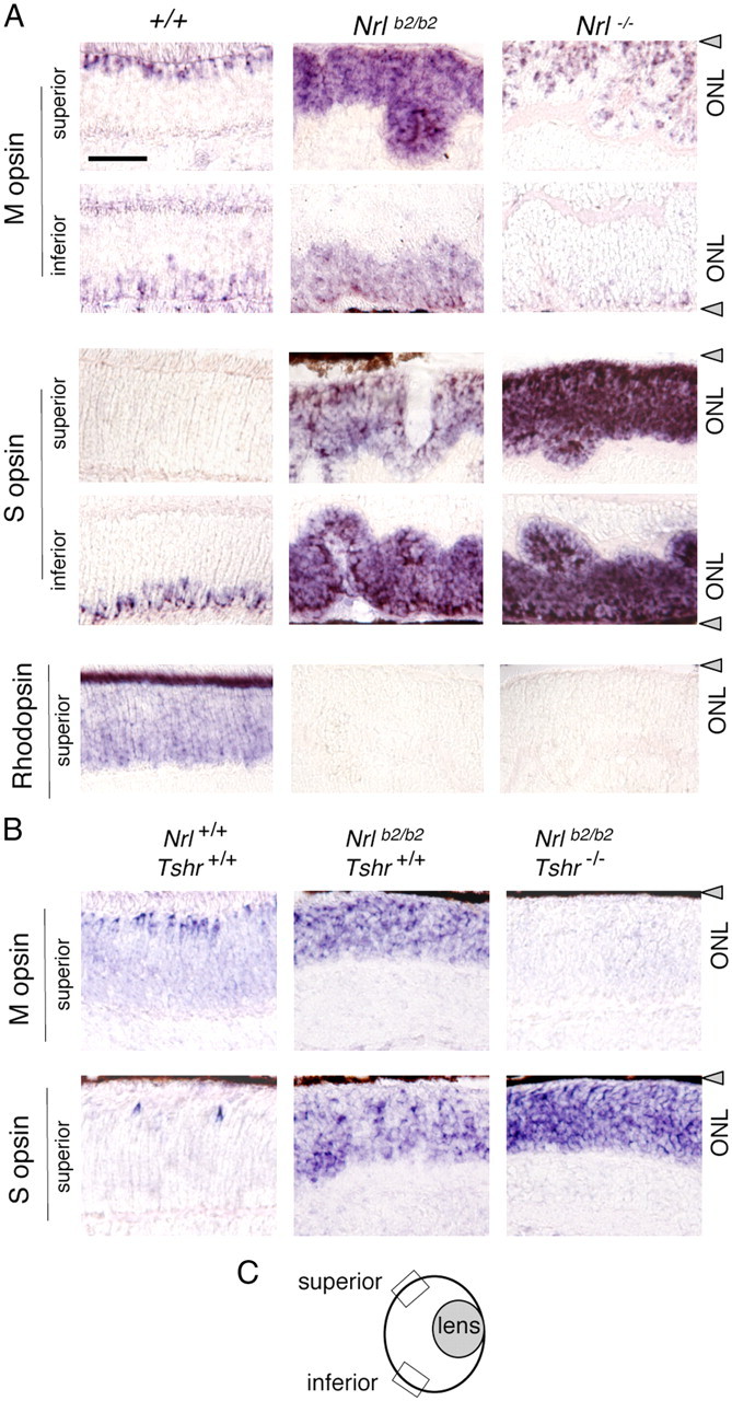
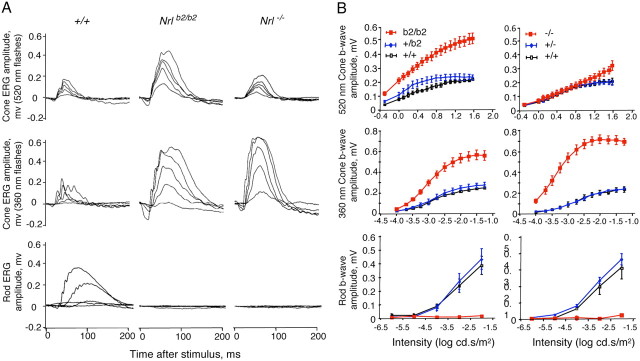
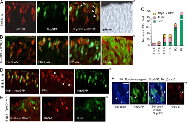
The typical mammalian visual system is based upon three photoreceptor types: rods for dim light vision and two types of cones (M and S) for color vision in daylight. However, the process that generates photoreceptor diversity and the cell type in which diversity arises remain unclear. Mice deleted for thyroid hormone receptor β2 (TRβ2) and neural retina leucine zipper factor (NRL) lack M cones and rods, respectively, but gain S cones. We therefore tested the hypothesis that NRL and TRβ2 direct a common precursor to a rod, M cone, or S cone outcome using Nrl(b2/b2) "knock-in" mice that express TRβ2 instead of NRL from the endogenous Nrl gene. Nrl(b2/b2) mice lacked rods and produced excess M cones in contrast to the excess S cones in Nrl(-/-) mice. Notably, the presence of both factors yielded rods in Nrl(+/b2) mice. The results demonstrate innate plasticity in postmitotic rod precursors that allows these cells to form three functional photoreceptor types in response to NRL or TRβ2. We also detected precursor cells in normal embryonic retina that transiently coexpressed Nrl and TRβ2, suggesting that some precursors may originate in a plastic state. The plasticity of the precursors revealed in Nrl(b2/b2) mice suggests that a two-step transcriptional switch can direct three photoreceptor fates: first, rod versus cone identity dictated by NRL, and second, if NRL fails to act, M versus S cone identity dictated by TRβ2.


Figures

Figure 1.
Figure 1.
Targeted replacement of Nrl gene with a Thrb2 gene. A, A Thrb2 cDNA, inserted at the start codon of Nrl, replaced both Nrl coding exons (gray boxes), creating the Nrlb2 allele. Filled triangle, Nrl gene promoter, open box, 5′-non-coding exon; TGA, stop codon; pA, polyadenylation site; loxP, residual loxP site after deletion of Neo selection gene. B, Western blot analysis showing expression of TRβ2 in Nrlb2/b2 mice. Arrowheads, specific bands for TRβ2 (∼58 kDa) and NRL (29–35 kDa). Thrb2−/− lane, TRβ2-deficient control at embryonic day 17.5 (E17.5). Numbers below lanes are signals quantified by densitometry relative to an arbitrary value of 1.0 in +/+ mice at the earliest age of detection; nd, not detectable. C, In situ hybridization showing ectopic expression of TRβ mRNA over the outer neuroblastic layer (ONBL) in Nrl+/b2 and Nrlb2/b2 mice at P7. Endogenous TRβ2 mRNA drops to low levels in the small cone population in +/+ neonates. Scale bar, 50 μm. INBL, Inner neuroblastic layer. RPE, Retinal pigmented epithelium (gray triangle).
Figure 2.
Figure 2.
Retinal histology in Nrl+/b2 and Nrlb2/b2 mice. A, Retinal histology in 3-month-old mice. The ONL in Nrl+/b2 mice contains cones (large nuclei with dispersed chromatin) and rods (smaller, denser nuclei) in the same proportions as +/+ mice. Nrlb2/b2 mice, like Nrl−/− mice, lack rods, have excess cones, and also display ONL folding and shortened outer segments (OS). INL, Inner nuclear layer; IPL, inner plexiform layer; IS, inner segments; OPL, outer plexiform layer. B, Higher magnification from images in A indicating cone nuclei (arrowheads) and a representative rod nucleus (r) in a +/+ mouse. Nrlb2/b2 mice possess only cone-like nuclei. Scale bars: A, B, 20 μm. C, Counts (mean ± SD) of cone and rod nuclei in ONL fields, determined on 3-μm-thick plastic sections like those in A. Fields avoided grossly folded areas in Nrlb2/b2 mice. Gray triangle, Retinal pigmented epithelium.
Figure 3.
Figure 3.
Distinct photoreceptor outcomes in Nrlb2/b2 and Nrl−/− mice. A, Western blot analysis of cone opsins and rhodopsin in retina from 6-week-old mice of the indicated Nrl genotypes. Thrb2−/− lane, Sample from TRβ2-deficient mice as a negative control for M opsin. B, Western blot analysis of cone opsins and TRβ2 in retina from P14 mice of the indicated Nrl genotypes. The initial induction of M opsin is pronounced in Nrlb2/b2 mice at this juvenile age. C, Quantitative PCR analysis of M opsin, S opsin, and TRβ2 mRNA levels in retina of mice of the indicated Nrl genotypes at P17.
Figure 4.
Figure 4.
Differential distribution of M and S opsins in Nrlb2/b2 and Nrl−/− mice. A, In situ hybridization analysis of cone opsin and rhodopsin mRNA in superior and inferior retinal regions in 8-week-old mice. Nrlb2/b2 mice overexpressed M and S opsins with a normal distribution trend, whereas in Nrl−/− mice, S opsin was strongly overexpressed in all retinal regions. Nrl−/− mice showed a small increase in the density of M opsin-positive cells in superior regions compared with +/+ mice, partly due to ONL folding in Nrl−/− mice. B, In situ hybridization analysis of opsin mRNA in hypothyroid Nrlb2/b2 mice at P17. On a hypothyroid Tshr−/− background, M opsin expression was retarded and S opsin overexpression exacerbated in Nrlb2/b2 mice. Scale bar: A (for A, B), 50 μm. C, Diagram of mouse eye section indicating location of superior and inferior retinal fields examined (boxes). Gray triangles, Retinal pigmented epithelium.
Figure 5.
Figure 5.
Distinct cone and rod functional responses in Nrl+/b2, Nrlb2/b2, and Nrl−/− mice. A, Representative electroretinogram traces for Nrlb2/b2 and Nrl−/− mice at ∼8 weeks of age. Top and middle rows, Photopic cone responses to stimuli with wavelengths of 520 and 360 nm that optimally activate mouse M and S opsins, respectively. Bottom row, Scotopic rod responses. Enhanced M and S cone responses were detected in Nrlb2/b2 mice, but only enhanced S cone responses in Nrl−/− mice. Families of traces are shown for light intensities of 0.5, 1.26, 2, 3.16, and 7.94 cd · s/m2 at 520 nm and 0.0001, 0.00032, 0.001, 0.0032 and 0.0316 cd · s/m2 at 360 nm for cones and 1 × 10⁻⁶, × 10−5, × 10−4, × 10−3, and × 10−2 cd · s/m2 for rods. B, Intensity–response curves of average ERG responses for groups of 4–6 mice (means ± SEM). Cone (top and middle rows) and rod (bottom row) ERG responses to varying stimulus intensities were determined for +/+, Nrl+/b2, and Nrlb2/b2 mice and, separately, for +/+, Nrl+/−, and Nrl−/− mice. Nrlb2/b2 and Nrl−/− mice were each compared with their own +/+ and heterozygous groups on comparable genetic backgrounds.
Figure 6.
Figure 6.
Transient coexpression of TRβ2 and Nrl in photoreceptor precursors. A, Double fluorescence analysis of cells positive for TRβ2 (@TRβ2 antibody, red), NrlpGFP (transgene, green), or both @TRβ2 and NrlpGFP (yellow or orange, arrows in merged image) in the outer neuroblastic layer of +/+ mouse retina at E16.5. Right, Phase contrast image of the same field. Confocal microscope images represent a single 1–1.5 μm z-plane obtained from 10-μm-thick mid-retinal cryosections (same in B–E). For orientation, the gray arrowhead indicates location of retinal pigmented epithelium. B, Double fluorescence analysis of cells positive for TRβ2 and NrlpGFP in +/+ mouse retina during development at E14.5, E18.5, P2, and P8. Doubly positive cells (yellow or orange) were most evident at E16.5–E18.5. Scale bars: A, B, 14 μm. C, Counts of cells positive for TRβ2, GFP, or both TRβ2 and GFP, determined on single z-plane confocal images from experiments shown in A and B. Counts shown were determined in mid-retinal fields. Counts in superior and inferior fields gave similar results. D, Verification of NrlpGFP transgene as a marker for cells expressing endogenous Nrl using an antibody against NRL protein (@Nrl) and direct fluorescence for NrlpGFP in +/+ embryos at E18.5. The @Nrl+ population included almost all GFP+ cells (yellowish and orange cells, merged image on the left) and a few @Nrl+ cells that were negative for GFP (white arrows, middle). E, Independent identification of cells that coexpress TRβ2 (@bGal) and endogenous NRL protein (@Nrl) (yellow or orange cells in merged image, left). Analysis was performed on E18.5 embryos homozygous for a targeted insertion of lacZ in the TRβ2-specific exon of the Thrb gene. Scale bars: D, E, 10 μm F, Immunofluorescence analysis for coexpression of S opsin (@S opsin antibody, blue), NrlpGFP (direct fluorescence, green) and Thrb2p-lacZ transgenes (@bGal antibody, red, indicator for TRβ2) in +/+ mice at P0. Arrows, S opsin/GFP doubly positive cell; arrowheads, S opsin/GFP/bGal triply positive cell; asterisks, S opsin-positive cell with no detectable GFP or bGal. Gray triangle, Retinal pigmented epithelium.

References

    1. Adler R, Raymond PA. Have we achieved a unified model of photoreceptor cell fate specification in vertebrates? Brain Res. 2008;1192:134–150. - PMC - PubMed
    1. Agathocleous M, Harris WA. From progenitors to differentiated cells in the vertebrate retina. Annu Rev Cell Dev Biol. 2009;25:45–69. - PubMed
    1. Akimoto M, Cheng H, Zhu D, Brzezinski JA, Khanna R, Filippova E, Oh EC, Jing Y, Linares JL, Brooks M, Zareparsi S, Mears AJ, Hero A, Glaser T, Swaroop A. Targeting of GFP to newborn rods by Nrl promoter and temporal expression profiling of flow-sorted photoreceptors. Proc Natl Acad Sci U S A. 2006;103:3890–3895. - PMC - PubMed
    1. Alfano G, Conte I, Caramico T, Avellino R, Arnò B, Pizzo MT, Tanimoto N, Beck SC, Huber G, Dollé P, Seeliger MW, Banfi S. Vax2 regulates retinoic acid distribution and cone opsin expression in the vertebrate eye. Development. 2011;138:261–271. - PubMed
    1. Applebury ML, Antoch MP, Baxter LC, Chun LL, Falk JD, Farhangfar F, Kage K, Krzystolik MG, Lyass LA, Robbins JT. The murine cone photoreceptor: a single cone type expresses both S and M opsins with retinal spatial patterning. Neuron. 2000;27:513–523. - PubMed

Publication types

MeSH terms