. 2011 Aug 3;31(31):11244-55.
doi: 10.1523/JNEUROSCI.1040-11.2011.

Extracellular signal-regulated kinase signaling in the ventral tegmental area mediates cocaine-induced synaptic plasticity and rewarding effects

Affiliations

Extracellular signal-regulated kinase signaling in the ventral tegmental area mediates cocaine-induced synaptic plasticity and rewarding effects

Bin Pan et al. J Neurosci. .

Abstract

Drugs of abuse such as cocaine induce long-term synaptic plasticity in the reward circuitry, which underlies the formation of drug-associated memories and addictive behavior. We reported previously that repeated cocaine exposure in vivo facilitates long-term potentiation (LTP) in dopamine neurons of the ventral tegmental area (VTA) by reducing the strength of GABAergic inhibition and that endocannabinoid-dependent long-term depression at inhibitory synapses (I-LTD) constitutes a mechanism for cocaine-induced reduction of GABAergic inhibition. The present study investigated the downstream signaling mechanisms and functional consequences of I-LTD in the VTA in the rat. Extracellular signal-regulated kinase (ERK) signaling has been implicated in long-term synaptic plasticity, associative learning, and drug addiction. We tested the hypothesis that VTA ERK activity is required for I-LTD and cocaine-induced long-term synaptic plasticity and behavioral effects. We show that the activation of receptors required for I-LTD increased ERK1/2 phosphorylation and inhibitors of ERK activation blocked I-LTD. We further demonstrate that ERK mediates cocaine-induced reduction of GABAergic inhibition and facilitation of LTP induction. Finally, we show that cocaine conditioned place preference (CPP) training (15 mg/kg; four pairings) increased ERK1/2 phosphorylation in the VTA, while bilateral intra-VTA injections of a CB(1) antagonist or an inhibitor of ERK activation attenuated ERK1/2 phosphorylation and the acquisition, but not the expression, of CPP to cocaine. Our study has identified the CB(1) and ERK signaling cascade as a key mediator of several forms of cocaine-induced synaptic plasticity and provided evidence linking long-term synaptic plasticity in the VTA to rewarding effects of cocaine.


Figures
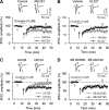
Figure 1.
Figure 1.
ERK, but not p38 MAPK, is required for eCB-dependent I-LTD in VTA dopamine neurons. A, The presence of cocaine (3 μm) 3 min before and 5 min during the 10 Hz stimulation [indicated by arrow (↑)] induced I-LTD in VTA dopamine neurons (n = 6). Bath application of the CB1 receptor antagonist AM251 (2 μm) blocked I-LTD (n = 7; p < 0.05 vs vehicle). Top, Sample IPSCs in the VTA before (indicated by “1”) and after (indicated by “2”) the 10 Hz stimulation. B, Bath application of MEK inhibitor SL327 (10 μm; n = 7) blocked I-LTD (p < 0.05 vs vehicle). C, MEK inhibitor U0126 (20 μm; n = 8) blocked I-LTD, whereas U0124 (20 μm; n = 7), the inactive analog of U0126, had no significant effect on I-LTD (p < 0.05 vs U0126). D, Bath application of p38 MAPK inhibitor SB 203580 (10 μm; n = 8) or SB 202190 (10 μm; n = 6) had no significant effects on I-LTD (p > 0.05 vs vehicle). For each group, six to eight neurons were recorded in six to eight slices that were prepared from four to six rats.
Figure 2.
Figure 2.
The CB1, D2, or group I mGluR agonists increased ERK1/2 phosphorylation but had no significant effect on p-38 phosphorylation in the VTA. A, VTA slices were treated for 10 min with vehicle (0.01% DMSO), the CB1 receptor agonist WIN 55212-2 (2 μm), the CB1 receptor antagonist AM251 (2 μm), or MEK inhibitor U0126 (20 μm). Quantitative group data (top) and representative Western blots (bottom) of phosphorylated ERK1/2 (p-ERK) in VTA homogenates are shown (n = 5 for each group; **p < 0.01, ***p < 0.001). B, Group I mGluR agonist DHPG (50 μm; 10 min) increased ERK1/2 phosphorylation; this effect was attenuated by AM251 and was abolished by U0126 (n = 4 for each group; **p < 0.01, ***p < 0.001). C, D2 receptor agonist quinpirole (10 μm; 10 min) increased ERK1/2 phosphorylation; this effect was not affected by AM251 but was abolished by U0126 (n = 5 for each group; **p < 0.01, ***p < 0.001). D, WIN 55212-2, DHPG, and quinpirole had no significant effects on p-p38 levels in VTA homogenates (n = 4 for each group; p > 0.05). For each group, four to five slices were prepared from three to four rats. Error bars indicate SEM.
Figure 3.
Figure 3.
Effects of in vivo pretreatment (intraperitoneal) with antagonists to receptors involved in I-LTD on cocaine-induced decrease in the amplitude of maximal IPSCs and mIPSCs in dopamine neurons in midbrain slices, which were prepared ∼24 h after the last saline or cocaine injection. A, Top, Sample IPSCs in response to stimuli of incremental intensities (20–140 μA). The neurons were voltage clamped at −20 mV. Bottom, Repeated intermittent intraperitoneal injections of cocaine for 5–7 d reduced the mean amplitude of maximal IPSCs compared with time-matched saline injections. Pretreatment (intraperitoneal) with CB1 receptor antagonist AM251 (2 mg/kg, i.p.) or MEK inhibitor SL327 (50 mg/kg, i.p.) 20 min before each intraperitoneal cocaine injection abolished cocaine-induced reduction of the mean amplitude of maximal IPSCs (n = 8–12 for each bar; **p < 0.01). B, Same as in A, except that mIPSCs were recorded at a holding potential of −70 mV (n = 7–10 for each bar; **p < 0.01). Sample mIPSCs from saline or cocaine-treated groups are shown on the top. In each group, 8–12 neurons were recorded in 8–12 slices, which were prepared from four to six rats. Error bars indicate SEM.
Figure 4.
Figure 4.
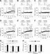
Effects of in vivo pretreatment (intraperitoneal) of AM251 or SL327 on cocaine-induced facilitation of LTP induction in VTA dopamine neurons. A, The EPSP–spike pairing protocol induced LTP in midbrain slices prepared from rats treated with vehicle plus cocaine for 5–7 d (n = 7), but not from rats treated with vehicle plus saline for 5–7 d (n = 8). The arrow indicates application of the LTP induction protocol. Experiments in A–C were performed in the absence of picrotoxin. B, C, Pretreatment (intraperitoneal) with CB1 receptor antagonist AM251 (2 mg/kg, i.p.) or MEK inhibitor SL327 (50 mg/kg, i.p.) 20 min before each intraperitoneal cocaine injection abolished cocaine-induced facilitation of LTP induction (n = 7–9). The same drug pretreatment had no significant effects on EPSPs in saline-treated rats (n = 7–8). D–F, The experiments are same in A–C, except that picrotoxin (50 μm) was included in the ACSF to block GABAergic inhibition (n = 6–9). G, H, Summary of LTP induction under various conditions in the absence (G) or presence (H) of picrotoxin. EPSP amplitude (percentage) was calculated as follows: 100 × (mean amplitude of EPSPs during the final 10 min of recording/mean amplitude of baseline EPSPs). *p < 0.05 compared with vehicle control. In each group, six to nine neurons were recorded in six to nine slices from four to five rats. Error bars indicate SEM.
Figure 5.
Figure 5.
The CB1 receptor activation contributes to cocaine-induced activation of ERK in the VTA. Immunostaining of activated ERK was performed using a phosphospecific antibody reacting with p-ERK. A, Intraperitoneal injection of AM251 (2 mg/kg) or SL327 (50 mg/kg) 20 min before each saline injection did not significantly affected the number of p-ERK-positive cells in the VTA. B, Repeated daily cocaine injections (15 mg/kg, i.p.; 6 d) increased p-ERK-positive cells in the VTA compared with repeated saline injections. Intraperitoneal injection of AM251 or SL327 20 min before each cocaine injection significantly attenuated cocaine-induced increase in the number of ERK-positive cells. Bottom, High-magnification view indicates that soma and dendrites of VTA neurons were labeled with p-ERK. Scale bars, 20 μm. C, Summarized data of the number of p-ERK-positive cells under various experimental conditions (4 rats each group; ***p < 0.001). Error bars indicate SEM.
Figure 6.
Figure 6.
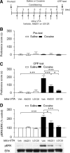
Intra-VTA infusions of the CB1 receptor antagonist AM251 or MEK inhibitor U0126 during the conditioning phase attenuated the acquisition of CPP to cocaine. A, Timeline of drug treatment and behavioral paradigm. Groups of rats received saline and cocaine place conditioning once daily for 8 d. Vehicle (Veh, 0.5 μl), AM251 (5 μg), or U0126 (0.1 μg) was bilaterally infused into the VTA 20 min before each cocaine or saline pairing. B, Pretest indicates that rats did not exhibit significant baseline bias in place preference in all groups (n = 6–9; p > 0.05). C, Intra-VTA infusions of AM251 or U0126 significantly attenuated CPP in cocaine-conditioned rats but did not affect CPP scores in saline-conditioned rats (n = 6–9; *p < 0.05, ***p < 0.001). D, ERK1/2 phosphorylation in the VTA was significantly increased in cocaine-conditioned rats compared with that in saline-conditioned rats. Intra-VTA infusions of AM251 or U0126 during the conditioning phase significantly decreased ERK1/2 phosphorylation (n = 4 for each group; *p < 0.01, ***p < 0.001). Western blotting was performed on VTA samples collected ∼1 h after the CPP tests. Error bars indicate SEM.
Figure 7.
Figure 7.
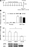
Intra-VTA infusions of AM251 or U0126 20 min before CPP test did not affect the expression of CPP to cocaine. A, Timeline of drug treatment and behavioral paradigm. Rats received only cocaine place conditioning. Vehicle (0.5 μl), AM251 (5 μg), or U0126 (0.1 μg) was bilaterally infused into the VTA 20 min before the CPP test. B, Cocaine conditioning induced significant increase in CPP scores compared with pretest. Intra-VTA infusion of AM251 or U0126 before the CPP test did not significantly affect CPP scores (n = 7–9; p > 0.05). C, Intra-VTA AM251 did not affect ERK1/2 phosphorylation in the VTA, whereas intra-VTA U0126 blocked ERK1/2 phosphorylation (n = 4 for each group; *p < 0.05). Western blotting was performed on VTA samples collected ∼1 h after the CPP tests. Error bars indicate SEM.
Figure 8.
Figure 8.
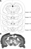
Histological verification of VTA cannula placements. A, Locations of intra-VTA infusion sites from the behavioral data summarized in Figures 6 and 7. For clarity, representative samples of bilateral cannula placements are shown. B, Cresyl violet-stained coronal section of typical bilateral intra-VTA cannula placements.

Similar articles

Cited by

References

    1. Argilli E, Sibley DR, Malenka RC, England PM, Bonci A. Mechanism and time course of cocaine-induced long-term potentiation in the ventral tegmental area. J Neurosci. 2008;28:9092–9100. - PMC - PubMed
    1. Atkins CM, Selcher JC, Petraitis JJ, Trzaskos JM, Sweatt JD. The MAPK cascade is required for mammalian associative learning. Nat Neurosci. 1998;1:602–609. - PubMed
    1. Berhow MT, Hiroi N, Nestler EJ. Regulation of ERK (extracellular signal-regulated kinase), part of the neurotrophin signal transduction cascade, in the rat mesolimbic dopamine system by chronic exposure to morphine or cocaine. J Neurosci. 1996;16:4707–4715. - PMC - PubMed
    1. Bolshakov VY, Carboni L, Cobb MH, Siegelbaum SA, Belardetti F. Dual MAP kinase pathways mediate opposing forms of long-term plasticity at CA3-CA1 synapses. Nat Neurosci. 2000;3:1107–1112. - PubMed
    1. Bouaboula M, Poinot-Chazel C, Bourrié B, Canat X, Calandra B, Rinaldi-Carmona M, Le Fur G, Casellas P. Activation of mitogen-activated protein kinases by stimulation of the central cannabinoid receptor CB1. Biochem J. 1995;312:637–641. - PMC - PubMed

Publication types

MeSH terms

LinkOut - more resources