. 2011 Aug 3;31(31):11282-94.
doi: 10.1523/JNEUROSCI.1810-11.2011.

Repeated amphetamine exposure disrupts dopaminergic modulation of amygdala-prefrontal circuitry and cognitive/emotional functioning

Affiliations

Repeated amphetamine exposure disrupts dopaminergic modulation of amygdala-prefrontal circuitry and cognitive/emotional functioning

Maric T L Tse et al. J Neurosci. .

Abstract

Repeated exposure to psychostimulants such as amphetamine (AMPH) disrupts cognitive and behavioral processes mediated by the medial prefrontal cortical (mPFC) and basolateral amygdala (BLA). The present study investigated the effects of repeated AMPH exposure on the neuromodulatory actions of dopamine (DA) on BLA-mPFC circuitry and cognitive/emotional processing mediated by these circuits. Rats received five AMPH (2 mg/kg) or saline injections (controls) over 10 d, followed by 2-4 week drug washout. In vivo neurophysiological extracellular recordings in urethane-anesthetized rats were used to obtain data from mPFC neurons that were either inhibited or excited by BLA stimulation. In controls, acute AMPH attenuated BLA-evoked inhibitory or excitatory responses; these effects were mimicked by selective D(2) or D(1) agonists, respectively. However, in AMPH-treated rats, the ability of these dopaminergic manipulations to modulate BLA-driven decreases/increases in mPFC activity was abolished. Repeated AMPH also blunted the excitatory effects of ventral tegmental area stimulation on mPFC neural firing. Behavioral studies assessed the effect of repeated AMPH on decision making with conditioned punishment, a process mediated by BLA-mPFC circuitry and mesocortical DA. These treatments impaired the ability of rats to use conditioned aversive stimuli (footshock-associated cue) to guide the direction of instrumental responding. Collectively, these data suggest that repeated AMPH exposure can lead to persistent disruption of dopaminergic modulation of BLA-mPFC circuitry, which may underlie impairments in cognitive/emotional processing observed in stimulant abusers. Furthermore, they suggest that impairments in decision making guided by aversive stimuli observed in stimulant abusers may be the result of repeated drug exposure.


Figures
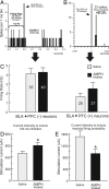
Figure 1.
Figure 1.
BLA stimulation can induce two distinct responses in separate populations of mPFC neurons (inhibitory and excitatory). Representative PSTH of BLA evoked inhibitory [BLA → PFC(−); A] and [excitatory BLA → PFC(+); B] responses. C, BLA → PFC(−) (left) and BLA → PFC(+) (right) neurons showed comparable basal firing rate between saline- and AMPH-treated rats. Numbers on each bar represent the n for each group. D, For BLA → PFC(−) neurons, higher stimulation currents were required to induce a comparable duration of inhibition in AMPH-treated rats relative to controls. E, Conversely, for BLA → PFC(+) cells, lower stimulation currents were required to induce maximal firing in AMPH-treated rats. ☆p < 0.05, between-group difference.
Figure 2.
Figure 2.
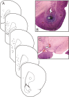
Histology. A, Schematic coronal slices of the mPFC showing representative locations in which BLA-evoked inhibitory (−) and excitatory (+) responses were observed. A displays photomicrographs of representative placements of stimulating electrodes at the BLA (B) and the VTA (C). Arrows highlight the location of the stimulating electrode tips.
Figure 3.
Figure 3.
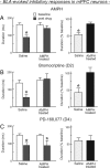
Changes in BLA-evoked inhibition induced by DA agonists. Left panels represent mean ± SEM duration of evoked inhibition under baseline conditions (white bars) and after treatment with DA agonists (gray bars); ☆p < 0.05, difference relative to baseline. Right panels are these data expressed as a percentage change from baseline; ☆p < 0.05, between-group difference. A, Treatment with AMPH (0.5 mg/kg, i.v.) significantly attenuates BLA-evoked inhibition in control rats (n = 10 cells, 10 rats), but this effect was abolished in rats that had received repeated AMPH (n = 10 cells, 10 rats). B, Treatment with the D2 receptor agonist bromocriptine (0.5 mg/kg, i.v.) also attenuated BLA-evoked inhibition in control rats (n = 5 cells, 5 rats) but not in AMPH treated rats (n = 6 cells, 6 rats), suggesting that these treatments induce a disruption in D2 receptor signaling. C, Treatment with the D4 receptor agonist PD 168,077 (1.0 mg/kg, i.v.) was effective in reducing BLA-evoked inhibition in both groups (n = 5 cells, 5 rats per group).
Figure 4.
Figure 4.
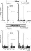
Representative PSTH illustrating the effect of D2 receptor stimulation on BLA → PFC(−) in control and AMPH-treated rats. Each histogram is compiled from firing data obtained over 150 single-pulse stimulations (sweeps) of the BLA. Stimulation of D2 receptors reduced the duration of inhibition induced by BLA stimulation in this neuron recorded from a saline-treated rat (top). However, similar treatments were ineffective at altering BLA-evoked inhibition in a neuron recorded from a rat that had received repeated AMPH (bottom).
Figure 5.
Figure 5.
Effects of repetitive burst stimulation of VTA on BLA-evoked inhibition of mPFC neurons. A, VTA stimulation leads to an attenuation of BLA-evoked inhibition in saline-treated (n = 8 cells, 4 rats) but not AMPH-treated rats (n = 10 cells, 7 rats). All conventions same as Figure 3. B, Repeated burst stimulation of the VTA also increased the spontaneous firing rate of these neurons in saline-treated but not AMPH-treated animals. ☆p < 0.05, between-group difference.
Figure 6.
Figure 6.
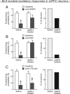
Changes in BLA-evoked firing induced by DA agonists and VTA stimulation. Left panels represent mean ± SEM evoked firing probability under baseline conditions (white bars) and after treatment with DA agonists/VTA stimulation (gray bars); ☆p < 0.05, difference relative to baseline. Right panels display the proportion of neurons recorded from saline- and AMPH-treated rats in which BLA-evoked firing was attenuated by these treatments. A, Treatment with AMPH (0.5 mg/kg, i.v.) attenuates BLA-evoked firing in controls (n = 9 cells, 9 rats), but this effect was not statistically reliable in rats receiving repeated AMPH treatments (n = 8 cells, 8 rats). B, Treatment with the D1 receptor agonist SKF81297 (0.5 mg/kg, i.v.) also attenuated BLA-evoked firing in control rats but not in AMPH-treated rats suggesting that these treatments induce a disruption in D1 receptor function (n = 6 cells, 6 rats per group). C, Suppression of evoked-firing induced by VTA burst stimulation did not differ between saline-treated (n = 6 cells, 4 rats) and AMPH-treated (n = 7 cells, 7 rats) animals.
Figure 7.
Figure 7.
Representative PSTH illustrating the effect of D1 receptor stimulation on BLA → PFC(+) in control and AMPH-treated rats. Each histogram is compiled from firing data obtained over 40 single-pulse stimulations of the BLA. Stimulation of D1 receptors with SKF81297 reduced spike firing evoked by BLA stimulation in a neuron recorded from a saline-treated rat (top). However, similar treatments were ineffective at altering BLA-evoked firing in a rat that had received repeated AMPH (bottom).
Figure 8.
Figure 8.
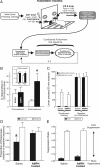
The effects of repeated AMPH treatment on decision making with conditioned punishment. A, Schematic of the training and testing schedule used for this experiment. After initial training to press two levers for food delivered on a VI 60 s schedule, rats were required to discriminate between one lever that delivered a compound conditioned stimuli (CS+) and footshock and another lever that delivered different compound stimulus (CS-0), each delivered on a VI 120 s schedule. After ∼25 d of training, they received repeated AMPH or saline treatments, a 7 d drug washout period, retraining with punishment, and then two test of conditioned punishment. B, After ∼25 d of initial punishment training, rats displayed a prominent bias away from the shock-associated lever (pretreatment). After repeated drug/saline treatment and drug washout, AMPH-treated rats displayed a significantly weaker bias than controls on the first day of punishment retraining (☆p < 0.05, between-group difference). Inset displays total number of lever presses made by rats in both groups before repeated AMPH/saline treatment (Pre) and on the first day of retraining after treatment (Post), which did not differ between groups. C, After several sessions of punishment retraining, both groups showed a strong bias toward the neural, CS-0-associated lever (white bars) relative to the aversive, shock-associated lever (black bars). This bias was not apparent when the CSs/shocks were omitted during baseline lever-pressing sessions, when rats only received food. D, During the critical tests of conditioned punishment, saline-treated rats again biased instrumental responding away from the lever producing aversive CS+ (now, without shocks). However, AMPH-treated rats showed no bias. E, Condition suppression of lever pressing (a measure of pavlovian fear) did not differ between the two groups. For D and E, ☆p < 0.05 aversive/CS+ versus neutral/CS-0.

References

    1. Au-Young SM, Shen H, Yang CR. Medial prefrontal cortical output neurons to the ventral tegmental area (VTA) and their responses to burst-patterned stimulation of the VTA: neuroanatomical and in vivo electrophysiological analyses. Synapse. 1999;34:245–255. - PubMed
    1. Bechara A, Dolan S, Denburg N, Hindes A, Anderson SW, Nathan PE. Decision-making deficits, linked to a dysfunctional ventromedial prefrontal cortex, revealed in alcohol and stimulant abusers. Neuropsychologia. 2001;39:376–389. - PubMed
    1. Bolles RC, Uhl CN, Wolfe M, Chase PB. Stimulus learning versus response learning in a discriminated punishment situation. Learn Motiv. 1975;6:439–444.
    1. Bowers MS, McFarland K, Lake RW, Peterson YK, Lapish CC, Gregory ML, Lanier SM, Kalivas PW. Activator of G protein signaling 3: a gatekeeper of cocaine sensitization and drug seeking. Neuron. 2004;42:269–281. - PMC - PubMed
    1. Briand LA, Flagel SB, Garcia-Fuster MJ, Watson SJ, Akil H, Sarter M, Robinson TE. Persistent alterations in cognitive function and prefrontal dopamine D2 reecptor following extended, but not limited, access to self-administered cocaine. Neuropsychopharmacology. 2008;33:2969–2980. - PMC - PubMed

Publication types

MeSH terms

LinkOut - more resources