. 2011 Oct;22(19):3634-44.
doi: 10.1091/mbc.E11-03-0197. Epub 2011 Aug 3.

Trs65p, a subunit of the Ypt1p GEF TRAPPII, interacts with the Arf1p exchange factor Gea2p to facilitate COPI-mediated vesicle traffic

Affiliations

Trs65p, a subunit of the Ypt1p GEF TRAPPII, interacts with the Arf1p exchange factor Gea2p to facilitate COPI-mediated vesicle traffic

Shuliang Chen et al. Mol Biol Cell. 2011 Oct.

Abstract

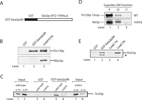
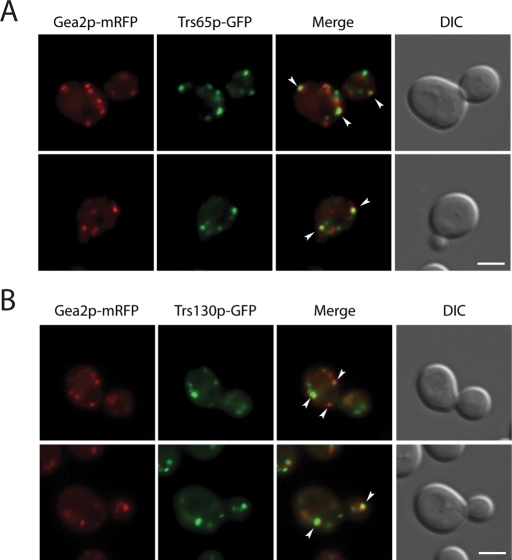
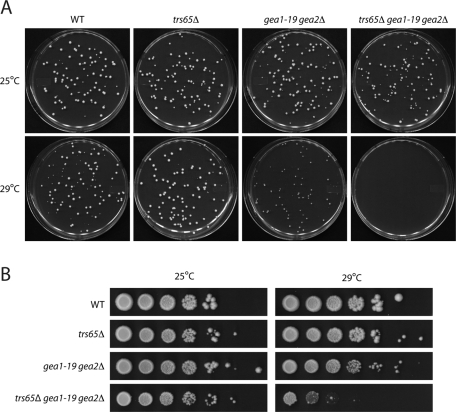
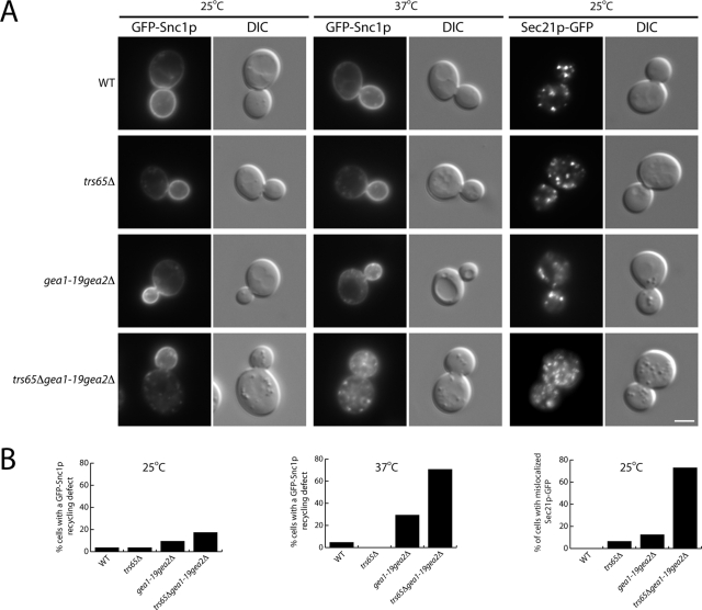
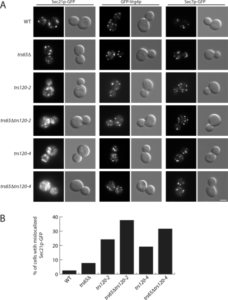
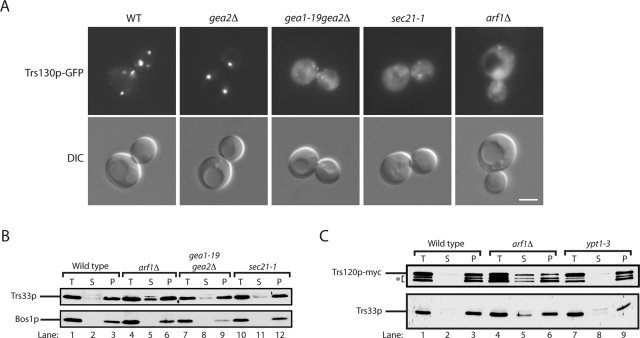
The TRAPP complexes are multimeric guanine exchange factors (GEFs) for the Rab GTPase Ypt1p. The three complexes (TRAPPI, TRAPPII, and TRAPPIII) share a core of common subunits required for GEF activity, as well as unique subunits (Trs130p, Trs120p, Trs85p, and Trs65p) that redirect the GEF from the endoplasmic reticulum-Golgi pathway to different cellular locations where TRAPP mediates distinct membrane trafficking events. Roles for three of the four unique TRAPP subunits have been described before; however, the role of the TRAPPII-specific subunit Trs65p has remained elusive. Here we demonstrate that Trs65p directly binds to the C-terminus of the Arf1p exchange factor Gea2p and provide in vivo evidence that this interaction is physiologically relevant. Gea2p and TRAPPII also bind to the yeast orthologue of the γ subunit of the COPI coat complex (Sec21p), a known Arf1p effector. These and previous findings reveal that TRAPPII is part of an Arf1p GEF-effector loop that appears to play a role in recruiting or stabilizing TRAPPII to membranes. In support of this proposal, we show that TRAPPII is more soluble in an arf1Δ mutant.


Figures

FIGURE 1:
FIGURE 1:
Two-hybrid analysis reveals that the N-terminus of Gea2p interacts with Cog4p, whereas the C-terminus of Gea2p interacts with Trs65p. A yeast two-hybrid screen was performed using either pGBKT7-Gea2N (amino acids 1–550) or pGBKT7-Gea2C (amino acids 759–1460) as bait and a yeast library in pACT2, as described in Materials and Methods. (A) Schematic diagram of yeast Gea2p. Cog4p was identified as an interacting partner of the N-terminal region of Gea2p, whereas Trs65p was identified as a partner for the C-terminal region. (B) Yeast strain AH109 was cotransformed with the indicated bait and prey plasmids. A dilution series of each transformant was spotted onto plates lacking leucine and tryptophan (−LEU−TRP) to select for the plasmids or onto medium lacking adenine and histidine (−ADE−HIS) to monitor expression of the ADE2 and HIS3 reporters. Plates were incubated at 30°C for 2 d. (C) Lysates were prepared as described in the Materials and Methods, incubated with glutathione Sepharose beads, washed, and blotted for Sec35p (subunit of the COG complex). (D) Same as C, except that the beads were blotted for Trs33p (subunit of the TRAPPII complex).
FIGURE 2:
FIGURE 2:
The C-terminus of Gea2p binds to TRAPPII. (A) Amino acids 972–1459 of Gea2p (Gea2pΔN) were fused to GST and expressed in bacteria. (B) Trs130p binds to the C-terminus of Gea2p. Purified GST (lane 1) and GST-Gea2pΔN (lane 2) were immobilized on glutathione Sepharose beads and incubated with lysate. Lane 3 contains 0.5% of the lysate that was used in the binding reaction. (C) Trs65p is required for the interaction of TRAPPII with Gea2p. GST (lanes 3 and 4) and GST-Gea2pΔN beads (lanes 5 and 6) were incubated with lysates prepared from wild type or cells in which the genomic copy of TRS65 was disrupted. The beads were washed and solubilized, and the solubilized sample was electrophoresed on an SDS–PAGE. Wild-type (lanes 1 and 2) and trs65Δ (lanes 7 and 8) lysates used in the binding experiment were subjected to SDS–PAGE and blotted for Trs33p. (D) The TRAPPII complex is largely intact in trs65Δ cells. Superdex-200 column fractions 9–11 prepared from wild-type (top) and trs65Δ cells (bottom) were probed for Trs120p-13myc and Bet3p. (E) TRAPPII binds directly to the C-terminus of Gea2p. Purified GST (lane 1) and GST-Gea2pΔN (lane 2) were immobilized on glutathione Sepharose beads and incubated with purified TAP-tagged TRAPP (lanes 1 and 2). The beads were washed and solubilized, and the solubilized sample was electrophoresed on a 12% SDS–PAGE. The gel was probed with the indicated antibodies. Lanes 3 and 4 contain 1 and 5%, respectively, of the purified TRAPP that was added to the binding reaction. The asterisk marks a breakdown fragment of Trs31p.
FIGURE 3:
FIGURE 3:
Gea2p partially colocalizes with TRAPPII. Gea2p partially colocalizes with the TRAPPII-specific subunits Trs65p (A) and Trs130p (B). Yeast cells expressing Trs65p-GFP and Gea2p-mRFP (A) or Trs130p-GFP and Gea2p-mRFP (B) were grown at 25°C in synthetic complete media to early log phase and examined by fluorescence microscopy. Scale bar, 3 μm.
FIGURE 4:
FIGURE 4:
The loss of TRS65 exaggerates the ts growth defect in the gea1-19gea2Δ mutant. (A) Yeast cells were grown at 25°C in YPD media to early stationary phase. Approximately 100 cells were then seeded on a YPD plate that was incubated at 25 or 29°C for 4 d. The concentration of cells was determined using a hemocytometer. (B) Cells were cultured at 25°C in YPD media to early stationary phase. After the cell concentration was adjusted to ∼1 × 10⁸ cells/ml, the cells were serially (10-fold) diluted and spotted onto YPD plates. The plates were incubated at 25 or 29°C for 4 d.
FIGURE 5:
FIGURE 5:
The loss of TRS65 exaggerates the Snc1p recycling defect and the COPI localization defect in the gea1-19gea2Δ mutant. (A) Yeast cells expressing GFP-Snc1p or Sec21p-GFP were grown in YPD media at 25°C to early log phase and pelleted. Two OD600 units of cells were harvested, shifted to prewarmed media at 37°C, and incubated for 5 min before the cells were directly examined by fluorescence microscopy. Scale bar, 3 μm. (B) Quantitation of the data in A. The data shown are representative. Approximately 34–46 cells were quantitated.
FIGURE 6:
FIGURE 6:
The loss of Trs65p exaggerates the COPI localization defect in the trs120-2 and trs120-4 mutants. (A) Yeast cells expressing Sec21p-GFP, GFP-Vrg4p or Sec7p-GFP were grown to early log phase in YPD medium at 25°C. A total of 0.4 OD600 unit of cells was harvested, resuspended in ice-cold YPD media, and directly examined by fluorescence microscopy. Scale bar, 3 μm. (B) Quantitation of data in A. The data shown are representative. For the trs120-2 and trs120-4 single and double mutants, ∼114–285 cells were quantitated.
FIGURE 7:
FIGURE 7:
TRAPPII is more soluble in the arf1Δ mutant. (A) Yeast cells expressing Trs130p-3xGFP were grown in YPD media at 25°C to early log phase and pelleted. Two OD600 units of cells were harvested, shifted to prewarmed media at 37°C, and incubated for 1 h before the cells were directly examined by fluorescence microscopy. Scale bar, 3 μm. (B) Differential fractionation experiments were performed as described in Materials and Methods, and Western blot analysis was performed. Top, the distribution of Trs33p; bottom, the distribution of the membrane protein Bos1p. (C) Same as B, except that the distribution of Trs120p-myc and Trs33p was monitored. The asterisk marks the breakdown of Trs120p-myc.

References

    1. Barrowman J, Bhandari D, Reinisch K, Ferro-Novick S. TRAPP complexes in membrane traffic: convergence through a common Rab. Nat Rev Mol Cell Biol. 2010;11:759–763. - PubMed
    1. Beck R, Adolf F, Weimer C, Bruegger B, Wieland FT. ArfGAP1 activity and COPI vesicle biogenesis. Traffic. 2009;10:307–315. - PubMed
    1. Bonifacino JS, Lippincott-Schwartz J. Coat proteins: shaping membrane transport. Nat Rev Mol Cell Biol. 2003;4:409–414. - PubMed
    1. Cai H, Reinisch K, Ferro-Novick S. Coats, tethers, Rabs, and SNAREs work together to mediate the intracellular destination of a transport vesicle. Dev Cell. 2007a;12:671–682. - PubMed
    1. Cai H, Yu S, Menon S, Cai Y, Lazarova D, Fu C, Reinisch K, Hay JC, Ferro-Novick S. TRAPPI tethers COPII vesicles by binding the coat subunit Sec23. Nature. 2007b;445:941–944. - PubMed

Publication types

MeSH terms

Substances

LinkOut - more resources