. 2011 Sep 1;187(5):2595-601.
doi: 10.4049/jimmunol.1100088. Epub 2011 Aug 3.

MPYS is required for IFN response factor 3 activation and type I IFN production in the response of cultured phagocytes to bacterial second messengers cyclic-di-AMP and cyclic-di-GMP

Affiliations

MPYS is required for IFN response factor 3 activation and type I IFN production in the response of cultured phagocytes to bacterial second messengers cyclic-di-AMP and cyclic-di-GMP

Lei Jin et al. J Immunol. .

Abstract

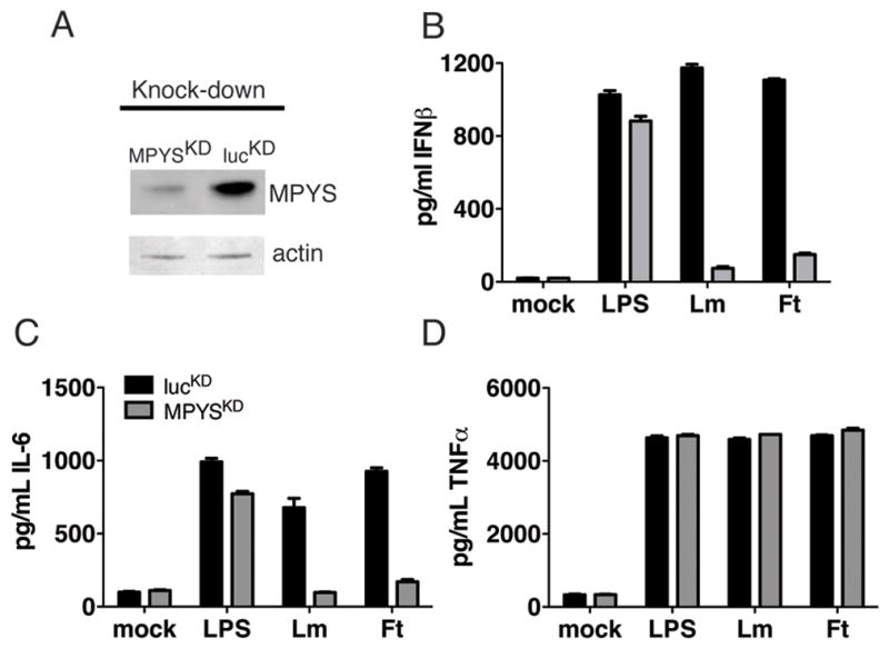
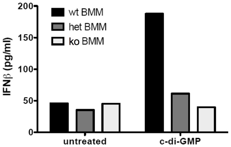
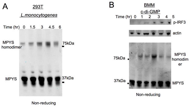
Cyclic-di-GMP and cyclic-di-AMP are second messengers produced by bacteria and influence bacterial cell survival, differentiation, colonization, biofilm formation, virulence, and bacteria-host interactions. In this study, we show that in both RAW264.7 macrophage cells and primary bone marrow-derived macrophages, the production of IFN-β and IL-6, but not TNF, in response to cyclic-di-AMP and cyclic-di-GMP requires MPYS (also known as STING, MITA, and TMEM173). Furthermore, expression of MPYS was required for IFN response factor 3 but not NF-κB activation in response to these bacterial metabolites. We also confirm that MPYS is required for type I IFN production by cultured macrophages infected with the intracellular pathogens Listeria monocytogenes and Francisella tularensis. However, during systemic infection with either pathogen, MPYS deficiency did not impact bacterial burdens in infected spleens. Serum IFN-β and IL-6 concentrations in the infected control and MPYS(-/-) mice were also similar at 24 h postinfection, suggesting that these pathogens stimulate MPYS-independent cytokine production during in vivo infection. Our findings indicate that bifurcating MPYS-dependent and -independent pathways mediate sensing of cytosolic bacterial infections.


Figures

Figure 1
Figure 1. MPYS is required for IFNβ production in response to infection with Listeria monocytogenes and Francisella tularensis.
A Whole Cell Lysate (WCL) from RAW264.7 cells expressing luciferase knockdown (lucKD) or MPYS-knockdown construct (MPYSKD) were fractionated by SDS- Polyacrylamide Gel Electrophoresis (PAGE) (10% NuPAGE), transferred to nitrocellulose and stained with anti-MPYS or anti-Actin Ab respectively. B–D. lucKD or MPYSKD macrophages were infected with Listeria monocytogenes (MOI 10) or Francisella tularensis LVS (MOI 100). Supernatants were collected at 12 hpi and ELISAs were performed for (B.) IFNβ, (C.) IL-6, and (D.) TNF.
Figure 2
Figure 2. MPYS is required for IFN β production in response to treatment with cyclic dinucleotides
A RAW264.7 lucKD or MPYSKD macrophages were treated with 9 μM c-di-AMP or 50 μM c-di-GMP and harvested for RNA at 12 hr post treatment. Quantitative PCR (TaqMan method) was performed for IFNβ gene expression and results were normalized to those of GAPDH. B. RAW264.7 lucKD or MPYSKD cells were treated with increasing concentrations of c-di-AMP or c-di-GMP as before. At 12 hr post treatment, supernatants were collected and analyzed by ELISA for IFNβ production.
Figure 3
Figure 3. MPYS is selectively required for IRF3 activation in response to treatment with cyclic dinucleotides
A Bone-marrow-derived macrophages (BMM) were treated with 20μM synthetic c-di-GMP or c-di-AMP as described in materials and methods. Whole cell lysates (WCL) were separated by SDS- PAGE (10% NuPAGE™), transferred to PVDF, and probed with Abs against p-IRF3 (Cell Signaling) and Actin (Santa Cruz). B. RAW264.7 lucKD or MPYSKD macrophages were treated with c-di-GMP (20μM) as before and harvested at the indicated times. WCL were separated and blotted as in A, then probed with Abs to p-IRF3, IκBα (Cell Signaling), actin or MPYS (34). C. The intensities of p-IRF3 (left panel) and IκBα (right panel) bands in B. were quantified using Odyssey 2.1 software and normalized to actin. The relative intensities of p-IRF3 and IκBα are plotted against time. D. RAW264.7 lucKD cells and MPYSKD cells were treated with c-di-AMP (20μM) as before. Activation of IRF3 and degradation of IκBα were evaluated as in B. E. The relative intensities of p-IRF3 and IκBα were determined as in C. Each experiment in this Figure has been done for at least three times.
Figure 4
Figure 4. Generation of MPYS-knockout mice by homologous recombination
A Strategy to generate MPYS-knock out mice. The genomic sequence of mouse MPYS gene is derived from a BAC clone RP24-490M12 (~140kb). MPYS gene consists of 8 exons and spans from 65182 to 72058bp in the BAC. Protein translation starts from exon 3. A hypothetic gene, 1700066B19Rik, is ~2.8kb downstream of MPYS and there is no protein-coding gene in the ~30kb region upstream of MPYS. The targeting construct covers ~10kb genomic region in the MPYS locus (64515~74175). The targeting construct has a neo gene inserted in the intron 5 and a diphtheria toxin gene at the 3 end of the MPYS gene. The neo gene is flanked by Frt elements and one LoxP site. Another LoxP site is inserted in the intron 2. Thus, using tissue specific Flp or Cre transgenic mice, we can also generate conditional KO or conditional WT MPYS mice. B. Western blot analysis of MPYS from WT, KO and heterozygous MPYS littermates. Splenocytes were lysed in RIPA buffer containing 0.1% SDS and run on a reducing SDS-PAGE gel. The blot was probed with rabbit α-MPYS Ab. N.S. non-specific. This experiment has been repeated more than three times. C. RT-PCR analysis of MPYS transcript from peripheral blood. RT-PCR was done using cDNA from peripheral blood cells of WT, KO and heterozygous MPYS littermates with primers for exon 2, 3, and 4 of the MPYS transcript. This experiment has been repeated twice.
Figure 5
Figure 5. MPYS-deficient BMM cells are defective for IFNβ production in response to treatment with cyclic dinucleotide monophosphates
Bone-marrow-derived macrophages (BMM) from indicated mice were treated with 50μM synthetic c-di-GMP as before. At 12 hr post treatment, supernatants were collected and analyzed by ELISA for IFNβ production. Experiments were repeated twice with similar results.
Figure 6
Figure 6. MPYS-deficient BMM cells fail to activate IRF3 in response to treatment with cyclic dinucleotide monophosphates
A–B Bone-marrow-derived macrophages (BMM) from indicated mice were treated with 20μM synthetic c-di-GMP (A.) or c-di-AMP (B.) for indicated times as before. WCL were separated by SDS-PAGE and probed with indicated Abs as in Fig 3. These experiments have been repeated twice.
Figure 7
Figure 7. MPYS forms homodimers in response to L. monocytogenes infection and cytosolic c-di-GMP activation
A. HEK-293T cells were infected with L. monocytogenes as in Fig. 1 for the indicated time. Cells were then lysed in RIPA buffer containing 0.1% SDS and fractionated using non-reducing SDS-PAGE gels. Blots were probed with the anti-MPYS Ab. B. BMM cells were treated with c-di-GMP (20μM) as in Fig. 3. Cells were then lysed and fractionated on a non-reducing gel. Blots were probed with indicated Abs. High molecular weight MPYS that previous studies have reported to be homodimers are indicated. These experiments have been repeated more than three times.
Figure 8
Figure 8. MPYS is required for early but not late IFN β or IL-6 productionin vivo
A–B. MPYS−/−, MPYS+/− and their WT littermates (B6) were infected with 10,000 cfu of L. monocytogenes (i.v) A. or 5,000 cfu F. tularensis (i.p.) B. Spleens were harvested, homogenized and dilution plated to determine bacterial burdens 72h (A.) or 48h (B.) later. Each point indicates an individual mouse. Bars indicate the mean values. C-F. MPYS−/−, MPYS+/− and their WT littermates (B6) (n=3) were infected as above with L. monocytogenes (C. & E.) or F. tularensis (D. & F.). Sera were collected at indicated times. IFNβ and IL-6 concentrations were measured by ELISA. Experiments were done twice.

References

    1. Serbina NV, Salazar-Mather TP, Biron CA, Kuziel WA, Pamer EG. TNF/iNOS-producing dendritic cells mediate innate immune defense against bacterial infection. Immunity. 2003;19:59–70. - PubMed
    1. Dinarello CA. Anti-cytokine therapeutics and infections. Vaccine. 2003;21(Suppl 2):S24–34. - PubMed
    1. Raychaudhuri SP, Nguyen CT, Raychaudhuri SK, Gershwin ME. Incidence and nature of infectious disease in patients treated with anti-TNF agents. Autoimmun Rev. 2009;9:67–81. - PubMed
    1. Decker T, Muller M, Stockinger S. The yin and yang of type I interferon activity in bacterial infection. Nat Rev Immunol. 2005;5:675–687. - PubMed
    1. Freudenberg MA, Merlin T, Kalis C, Chvatchko Y, Stubig H, Galanos C. Cutting edge: a murine, IL-12-independent pathway of IFN-gamma induction by gram-negative bacteria based on STAT4 activation by Type I IFN and IL-18 signaling. J Immunol. 2002;169:1665–1668. - PubMed

Publication types

MeSH terms