

Longitudinal changes of structural connectivity in traumatic axonal injury
- PMID: 21813787
- PMCID: PMC3162636
- DOI: 10.1212/WNL.0b013e31822c61d7
Longitudinal changes of structural connectivity in traumatic axonal injury
Abstract
Objectives: To identify structural connectivity change occurring during the first 6 months after traumatic brain injury and to evaluate the utility of diffusion tensor tractography for predicting long-term outcome.
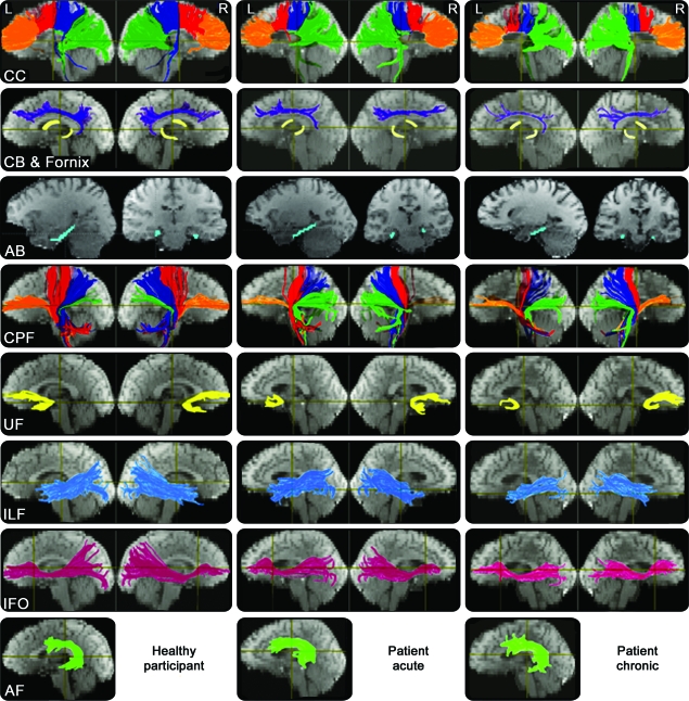
Methods: The participants were 28 patients with mild to severe traumatic axonal injury and 20 age- and sex-matched healthy control subjects. Neuroimaging was obtained 0-9 days postinjury for acute scans and 6-14 months postinjury for chronic scans. Long-term outcome was evaluated on the day of the chronic scan. Twenty-eight fiber regions of 9 major white matter structures were reconstructed, and reliable tractography measurements were determined and used.
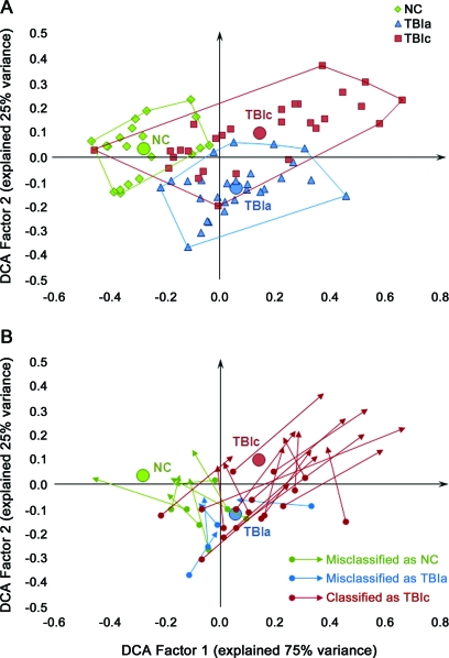
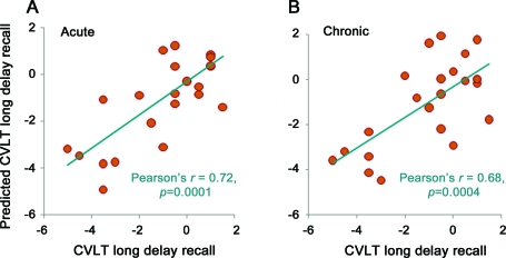
Results: Although most (23 of 28) patients had severe brain injury, their long-term outcome ranged from good recovery (16 patients) to moderately (5 patients) and severely disabled (7 patients). In concordance with the diverse outcome, the white matter change in patients was heterogeneous, ranging from improved structural connectivity, through no change, to deteriorated connectivity. At the group level, all 9 fiber tracts deteriorated significantly with 7 (corpus callosum, cingulum, angular bundle, cerebral peduncular fibers, uncinate fasciculus, and inferior longitudinal and fronto-occipital fasciculi) showing structural damage acutely and 2 (fornix body and left arcuate fasciculus) chronically. Importantly, the amount of change in tractography measurements correlated with patients' long-term outcome. Acute tractography measurements were able to predict patients' learning and memory performance; chronic measurements also determined performance on processing speed and executive function.
Conclusions: Diffusion tensor tractography is a valuable tool for identifying structural connectivity changes occurring between the acute and chronic stages of traumatic brain injury and for predicting patients' long-term outcome.
Figures
Comment in
-
White matter in traumatic brain injury: Dis- or dysconnection?Neurology. 2011 Aug 30;77(9):810-1. doi: 10.1212/WNL.0b013e31822b015b. Epub 2011 Aug 3. Neurology. 2011. PMID: 21813789 No abstract available.
References
-
- Graham DI, McIntosh TK, Maxwell WL, Nicoll JA. Recent advances in neurotrauma. J Neuropathol Exp Neurol 2000;59:641–651 - PubMed
-
- Gaetz M. The neurophysiology of brain injury. Clin Neurophysiol 2004;115:4–18 - PubMed
-
- Povlishock JT, Katz DI. Update of neuropathology and neurological recovery after traumatic brain injury. J Head Trauma Rehabil 2005;20:76–94 - PubMed
-
- Basser PJ, Mattiello J, LeBihan D. Estimation of the effective self-diffusion tensor from the NMR spin echo. J Magn Reson B 1994;103:247–254 - PubMed
-
- Basser PJ, Pierpaoli C. Microstructural and physiological features of tissues elucidated by quantitative-diffusion-tensor MRI. J Magn Reson B 1996;111:209–219 - PubMed
Publication types
MeSH terms
Grants and funding
LinkOut - more resources
Full Text Sources