. 2011 Oct 15;12(8):756-64.
doi: 10.4161/cbt.12.8.17169. Epub 2011 Oct 15.

DNA mismatch repair proficiency executing 5-fluorouracil cytotoxicity in colorectal cancer cells

Affiliations

DNA mismatch repair proficiency executing 5-fluorouracil cytotoxicity in colorectal cancer cells

Moriya Iwaizumi et al. Cancer Biol Ther. .

Abstract

Background: 5-fluorouracil (5FU)-based chemotherapy is the standard treatment for advanced stage colorectal cancer (CRC) patients. Several groups including ours have reported that stage II-III colorectal cancer patients whose tumors retain DNA Mismatch repair (MMR) function derive a benefit from 5FU, but patients with tumors that lost MMR function do not. Although MMR recognition of 5FU incorporated in DNA has been demonstrated biochemically, it has not been demonstrated within cells to execute 5FU cytotoxicity.

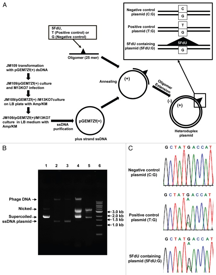
Aim: To establish an efficient construction model for 5FU within DNA and demonstrate that 5FU incorporated into DNA can trigger cellular cytotoxicity executed by the DNA MMR system.

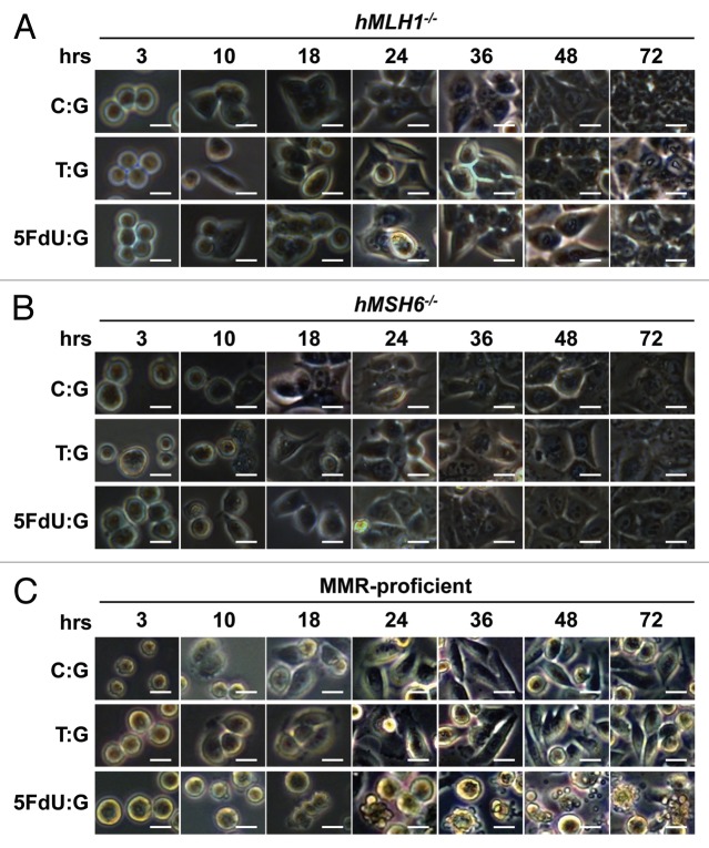
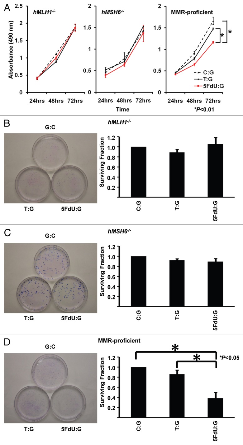
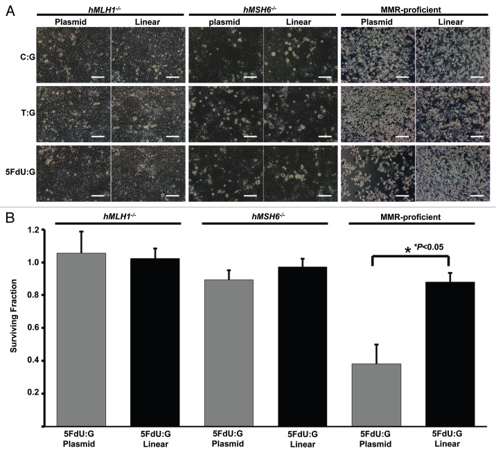
Methods: We constructed a 5FdU-containing heteroduplex plasmid (5FdU plasmid) and 5FdU-containing linear dsDNA (5FdU linear DNA), and transfected these into MMR-proficient, hMLH1-/- and hMSH6-/- cells. We observed cell growth characteristics of both transfectants for 5FU-induced cytotoxicity.

Results: MMR- proficient cells transfected with the 5FdU plasmid but not the 5FdU linear DNA showed reduced cell proliferation by MTS and clonogenic assays, and demonstrated cell morphological change consistent with apoptosis. In MMR-deficient cells, neither the 5FdU plasmid nor 5FdU linear DNA induced cell growth or morphological changes different from controls.

Conclusion: 5FdU as heteroduplex DNA in plasmid but not linear form triggered cytotoxicity in a MMR-dependent manner. Thus 5FU incorporated into DNA, separated from its effects on RNA, can be recognized by DNA MMR to trigger cell death.


Figures

Figure 1
Figure 1
Efficient construction of 5FdU containing heteroduplex plasmid. (A) Flowchart of for preparation of the 5FdU containing heteroduplex plasmid. (B) Electrophoretic analysis of various DNA preparations. Lane 1; parental pGEM7Zf(+) dsDNA plasmid isolated by an alkaline lysis method. Almost of all the plasmid was in supercoiled form. Lane 2; single-stranded DNA plasmid generated by conventional M13KO7 method (17). Lane 3; Single-stranded DNA isolated from JM109/pGEM7Zf(+)/M13KO7 complex. Lane 4; 5FdU-containing heteroduplex plasmid constructed by ssDNA plasmid generated by conventional M13KO7 method. Lane 5; 5FdU-containing heteroduplex plasmid constructed by ssDNA plasmid isolated from JM109/pGEM7Zf(+)/M13KO7 complex. Lane 6; linear dsDNA marker (1 kb Ladder N3232S, New England Biolabs). We generated less contaminated ssDNA by the bacteria/phagemid/helper phage complex method than by the conventional M13KO7 infection method (17) (lane 2 and 3), which also reduced the amount of contamination after the oligomer extension reaction (lane 4 and 5). (C) Sequence at the heteroduplex site. PCR was performed using the constructed plasmid as a template. After purifying the PCR product, direct sequencing was performed to confirm the heteroduplex site. KM, kanamycin; Amp, ampicillin; Nicked, nicked dsDNA plasmid; Supercoiled, supercoiled plasmid.
Figure 2
Figure 2
5FdU-containing heteroduplex plasmid leads to cellular morphological changes in MMR-proficient cells. Twelve hours after transfection, hMLH1-/- (A), hMSH6-/- (B) and MMR-proficient (C) cells were seeded at a density of 1.5 × 10⁵ cells per well onto 6-well plates and observed by microscope. By 24 h after seeding, the 5FdU plasmid induced morphological changes consistent with cell death in the MMR-proficient cells (C), while no morphological changes were observed in the hMLH1-/- (A) and hMSH6-/- (B) cells. Neither MMR-proficient cells nor MMR-deficient cells demonstrated morphological changes by heteroduplex (T:G) and negative (C:G) control plasmids. Scale bars; 10 µm. 5FdU:G; 5FdU containing heteroduplex plasmid, T:G; positive control heteroduplex plasmid, C:G; unaltered plasmid (negative control).
Figure 3
Figure 3
5FdU containing heteroduplex plasmid induces cytotoxicity in MMR-proficient cells. (A) MTS assay. Cells (5,000 cells/well) of each population were seeded in 100 µl of culture medium in the wells of three 96-well plastic plates and plates were incubated in a 5% CO2/95% air incubator for 24 , 48 and 72 h. After incubation, 10 µl of MTS reagent solution was added to each well, and the plate was incubated for an additional 4 h in the 5% CO2/95% air incubator. Cell viability was measured by scanning with a microplate reader at 490 nm. This experiment was performed three times. (B–D) Clonogenic assay. Twelve hours after transfection, hMLH1-/- (B), hMSH6-/- (C) and MMR-proficient (D) cells were seeded at a density of 5 × 10² cells/100 mm dish and colonies were stained with Giemsa and counted 14 days after seeding. Survival fraction was shown as a percentage of negative controls. This experiment was performed at least three times.
Figure 4
Figure 4
5FdU-containing heteroduplex plasmid is more useful for measurement of cytotoxicity than linear dsDNA. (A) Cell density. Twelve h after transfection, cells were seeded at a density of 1.5 × 10⁵ cells per well into 6-well plates. 72 h after seeding, the cell density was observed by microscopy. 5FdU-plasmid transfected MMR-proficient cells showed lower cell density than 5FdU linear dsDNA transfected cells. (B) Clonogenic assay. Twelve hours after transfection, cells were seeded at a density of 5 × 10² cells/100 mm dish and colonies were stained with Giemsa and counted 14 days after seeding. Survival fraction was shown as a percentage of negative controls. This experiment was performed at least three times. Scale bars; 100 µm.

References

    1. Laurie JA, Moertel CG, Fleming TR, Wieand HS, Leigh JE, Rubin J, et al. Surgical adjuvant therapy of large-bowel carcinoma: an evaluation of levamisole and the combination of levamisole and fluorouracil. The North Central Cancer Treatment Group and the Mayo Clinic. J Clin Oncol. 1989;7:1447–1456. - PubMed
    1. Moertel CG, Fleming TR, Macdonald JS, Haller DG, Laurie JA, Goodman PJ, et al. Levamisole and fluorouracil for adjuvant therapy of resected colon carcinoma. N Engl J Med. 1990;322:352–358. doi: 10.1056/NEJM199002083220602. - DOI - PubMed
    1. Moertel CG, Fleming TR, Macdonald JS, Haller DG, Laurie JA, Tangen CM, et al. Fluorouracil plus levamisole as effective adjuvant therapy after resection of stage III colon carcinoma: a final report. Ann Intern Med. 1995;122:321–326. - PubMed
    1. Boland CR, Sinicrope FA, Brenner DE, Carethers JM. Colorectal cancer prevention and treatment. Gastroenterology. 2000;118:115–128. doi: 10.1016/S0016-5085(00)70010-2. - DOI - PubMed
    1. Efficacy of intravenous continuous infusion of fluorouracil compared with bolus administration in advanced colorectal cancer. Meta-analysis Group In Cancer. J Clin Oncol. 1998;16:301–308. - PubMed

Publication types

MeSH terms