. 2011 Jul;9(7):e1001110.
doi: 10.1371/journal.pbio.1001110. Epub 2011 Jul 26.

Antagonism of LIN-17/Frizzled and LIN-18/Ryk in nematode vulva induction reveals evolutionary alterations in core developmental pathways

Affiliations

Antagonism of LIN-17/Frizzled and LIN-18/Ryk in nematode vulva induction reveals evolutionary alterations in core developmental pathways

Xiaoyue Wang et al. PLoS Biol. 2011 Jul.

Abstract

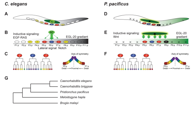
Most diversity in animals and plants results from the modification of already existing structures. Many organ systems, for example, are permanently modified during evolution to create developmental and morphological diversity, but little is known about the evolution of the underlying developmental mechanisms. The theory of developmental systems drift proposes that the development of conserved morphological structures can involve large-scale modifications in their regulatory mechanisms. We test this hypothesis by comparing vulva induction in two genetically tractable nematodes, Caenorhabditis elegans and Pristionchus pacificus. Previous work indicated that the vulva is induced by epidermal growth factor (EGF)/RAS and WNT signaling in Caenorhabditis and Pristionchus, respectively. Here, we show that the evolution of vulva induction involves major molecular alterations and that this shift of signaling pathways involves a novel wiring of WNT signaling and the acquisition of novel domains in otherwise conserved receptors in Pristionchus vulva induction. First, Ppa-LIN-17/Frizzled acts as an antagonist of WNT signaling and suppresses the ligand Ppa-EGL-20 by ligand sequestration. Second, Ppa-LIN-18/Ryk transmits WNT signaling and requires inhibitory SH3 domain binding motifs, unknown from Cel-LIN-18/Ryk. Third, Ppa-LIN-18/Ryk signaling involves Axin and β-catenin and Ppa-axl-1/Axin is epistatic to Ppa-lin-18/Ryk. These results confirm developmental system drift as an important theory for the evolution of organ systems and they highlight the significance of protein modularity in signal transduction and the dynamics of signaling networks.


Conflict of interest statement

The authors have declared that no competing interests exist.

Figures

Figure 1
Figure 1. Comparison of cell fate specification in the ventral epidermis in C. elegans and P. pacificus.
(A–C) Vulva formation in C. elegans. (A) P(3–8).p form the vulva equivalence group, P(1,2,9–11).p fuse with the hypodermal syncytium hyp7 (white circles). (B) P(5–7).p adopt a 2°–1°–2° vulval fate pattern (blue and red ovals), while P(3,4,8).p adopt an epidermal fate (3°, yellow ovals). The specification of vulval cell fates depends on LIN-3/EGF signaling, which originates from the gonadal AC (green arrow). P6.p signals its neighbors to adopt a 2° fate via LIN-12/Notch signaling (red arrow). EGL-20/Wnt is expressed as gradient from the posterior tail (grey) and establishes ground polarity of P5.p and P7.p. (C) Cell lineage and cell arrangement of P(5–7).p and their progeny. P6.p divides symmetrically to produce eight progeny that form the inner part of the vulva. P5.p and P7.p divide asymmetrically to produce seven progeny and form the anterior and posterior part of the vulva, respectively. The distal most progeny of P(5,7).p adhere to the epidermis. (D–F) Vulva formation in P. pacificus. (D) P(5–7).p adopted a 2°–1°–2° cell fate pattern, P(1–4, 9–11).p die of programmed cell death (black cross). P8.p does not divide but influences the fate of P(5,7).p (4° fate, black oval). (E) P. pacificus vulva induction depends on Wnt signaling and Wnt ligands are expressed in the somatic gonad (green arrow) and in the case of Ppa-EGL-20 in the posterior tail (green gradient). (F) Cell lineage and cell arrangement of P(5–7).p and their progeny. P6.p divides symmetrically and produces six progeny, whereas P(5,7).p have a cell lineage similar to C. elegans. (G) Schematic of the phylogenetic relationship of five nematodes with fully sequenced genomes. The shown distances do not represent the real divergence time for species. For P. pacificus and C. elegans, a divergence of 250–420 million y has been suggested on the basis of the sequence comparison of more than 1,000 orthologous genes .
Figure 2
Figure 2. Characterization of Ppa-lin-17(tu383) and analysis of its SDBMs.
(A) Nomarski photomicrograph of an early J4 larval stage of P. pacificus with an undifferentiated P8.p cell (white arrow). (B) Corresponding stage of a Ppa-lin-17(tu383) mutant with a vulva-like structure from P8.p (white arrow). (C) Mapping of tu383. Markers used for mapping tu383 to a small region on Chromosome V between markers L88.4 and L88.60 (black arrows). (D) Rescue of Ppa-lin-17(tu383). A 16-kb genomic DNA construct containing a wild-type copy of Ppa-lin-17 was coinjected with Ppa-egl-20::TurboRFP (as marker, red expression in the tail). White arrow points to P8.p, which did not differentiate in transgenic worms. (E) Cloning of Ppa-lin-17(tu383). The intracellular domain of Ppa-lin-17(tu383) and conceptual translation are shown. Ppa-lin-17(tu383) alters the stop codon (red letters), resulting in a 17 amino acid extension of the protein. The yellow boxes represent the repeated SDBMs, followed by a potential phosphorylation site. (F) Analysis of functional motifs of Ppa-lin-17(tu383). Frequency of vulva induction in P(1–4).p and P8.p were examined in a Ppa-ced-3(tu54) mutant background. Transgenic animals carry either a full length Ppa-lin-17(tu383) (blue bars), a SDBM-mutated form of Ppa-lin-17(tu383) (green, red, and orange bars), or a wild-type Ppa-lin-17 (light blue bars).
Figure 3
Figure 3. Molecular structure and function of Ppa-lin-18 and analysis of SDBMs in Ppa-LIN-18.
(A) Left, Ppa-LIN-18 protein structure. Wnt-inhibitory factor-1 like domain (WIF domain, white), Receptor related to tyrosine kinase (PTK_Ryk, pink) and transmembrane domain (TM, blue). Positions of three potential SDBMs of Ppa-LIN-18 are indicated by arrows. Right, amino acid sequence alignment of Ppa-LIN-18 and Cel-LIN-18. The positions of SDBMs in Ppa-LIN-18 are shown by black letters, the single potential SDBM in Cel-LIN-18 is shown in pink. (B) Nomarski photomicrograph of a J4 larval stage of a Ppa-mom-2(tu63); Ppa-lin-18(tu359) double mutant with a vulvaless phenotype. Black arrows point to the undifferentiated VPCs. (C) Corresponding stage of a transgenic animal carrying a wild-type copy of Ppa-LIN-18. P(5–7).p differentiated and formed normal vulva, P8.p remains epidermal (black arrows). (D) Rescue efficiency of Ppa-mom-2(tu363); Ppa-lin-18(tu359) by different Ppa-LIN-18 constructs (signal peptide, green; WIF domain, white; TM domain, blue; PTK_Ryk domain, pink). The WT-LIN-18-HA construct contains an HA epitope tag, “MYPYDVPDYA” (red) in front of the stop codon of Ppa-LIN-18.
Figure 4
Figure 4. Chimeric Ppa-LIN-17(tu383)-LIN-18 (SDBM) transgenes cause ectopic vulva formation.
(A) Design of chimeric constructs of Ppa-LIN-17(tu383)-LIN-18 (SDBM). The ectopic SDBM site of Ppa-LIN-17(tu383) was substituted by the SDBMs of Ppa-LIN-18 one by one. (B, C) Graphical (B) and numerical (C) analysis of vulva induction after overexpression of chimeric constructs in a Ppa-ced-3(tu54) background. Experimental design is similar to experiments in Figure 3F. Purple and orange bars show vulva differentiation in transgenic animals carrying chimeric constructs of Ppa-LIN-17(tu383) substitutions with the first and third SDBM of Ppa-LIN-18. In contrast, substitution of Ppa-LIN-17(tu383) with the second SDBM of Ppa-LIN-18 substitution showed limited vulva differentiation in P(1–4).p and P8.p. Statistic analysis: a p-value = 4.575e–06; b p-value = 0.0003; c p-value = 0.72; d p-value = 1; e p-value = 6.069e–07; f p-value = 0.001 (comparison with ced-3(tu54); tuEx26 [lin-17(tu383)PGIP574-577AAAA], Fisher exact test).
Figure 5
Figure 5. Molecular cloning and characterization of Ppa-axl-1.
(A) Vulval patterning defects of tu98 and tu329 alleles. Besides P(5–7).p, P8.p and often the surviving posterior cells P(9–11).p form vulva-like structures. *Average number of Pn.p cells with vulva-like differentiation is above 5. We did not count differentiation of P(10,11).p in the posterior region as it is known that these cells also form vulva-like protrusions in a ced-3 mutant background. (B) Nomarski photomicrograph of an early J4 larval stage of a Ppa-axl-1(tu98) mutant animal with a vulva-like structure in P8.p and P9.p (white arrows). (C) Nomarski photomicrograph of a late J4 larval stage of a Ppa-axl-1(tu98);Ppa-ced-3(tu54) double mutant with ectopic vulva-like structures in the anterior and posterior body region (black arrows). (D) Mapping of tu98 and tu329 to an interval on Chromosome V between markers S240 and S327 (black arrows). (E) Domain structure and position of mutations in Ppa-axl-1. Ppa-AXL-1 is most similar in sequence to Cel-AXL-1 and contains a RGS and the “Dishevelled-Axin” (DIX) domain. Mutations in both alleles are in the RGS domain. tu98 was induced by trimethylpsoralen/UV light treatment and contains two mutations, a phenomenon known to occur after this type of treatment. Phylogenetic tree of AXIN encoding genes. Branch length is proportional to the number of substitutions per site.
Figure 6
Figure 6. Expression of Ppa-lin-17 and Ppa-egl-20 control vulva induction in P. pacificus.
(A–C) J2 larval stage of wild-type P. pacificus animal. (A) Ppa-lin-17 promoter driving nuclear localized TurboRFP expression in the posterior tail. (B) Within the same animal, GFP under the control of the Ppa-egl-20 promoter. (C) Merged image of Ppa-egl-20::GFP; Ppa-lin-17::TurboRFP. (D) Ligand sequestration model. In wild type, Ppa-LIN-17 sequesters Ppa-EGL-20 in the posterior body region. In Ppa-lin-17 mutant animals, excessive Ppa-EGL-20 ligand reaches the central body region induces vulva formation in the absence of the signal from the second signaling center, the somatic gonad. A mutation in Ppa-egl-20 fully suppresses the gonad-independent vulva differentiation phenotype of Ppa-lin-17.
Figure 7
Figure 7. Model for the antagonism of Ppa-LIN-17 and Ppa-LIN-18 during Ppa-EGL-20 signaling.
(A) Vulva induction in wild type. Left, the first and third SDBM sites in Ppa-LIN-18 bind to an inhibitor (I), which in the absence of Wnt ligands prevents signal transduction. Right, Ppa-EGL-20 acts as one of the inducers of P. pacificus vulva formation. When the amount of Ppa-EGL-20 outcompetes ligand sequestration by Ppa-LIN-17, the inhibitor is released from Ppa-LIN-18 and signaling occurs. This Ppa-LIN-18/Ryk signaling involves Axin and β-catenin. (B) Vulva induction in Ppa-lin-17(tu383) mutant conditions. Left, the ectopic SDBM site of Ppa-lin-17(tu383) attracts the inhibitor and causes the release from Ppa-LIN-18, resulting in ligand-independent vulva differentiation. Consistently, the Ppa-lin-17(tu383) mutant phenotype is not suppressed by Ppa-egl-20 mutants.

Comment in

Similar articles

Cited by

References

    1. Raff R. A. The shape of life: genes, development, and the evolution of animal form. Chicago: The University of Chicago Press; 1996.
    1. Bento G, Ogawa A, Sommer R. J. Co-option of the hormone-signaling module dafachronic acid-DAF-12 in nematode evolution. Nature. 2010;466:494–497. - PubMed
    1. Schmidt-Rhaesa A. The evolution of organ systems. New York: Oxford University Press; 2007.
    1. True J. R, Haag E. S. Developmental system drift and flexibility in evolutionary trajectories. Evol Dev. 2001;3:109–119. - PubMed
    1. Sommer R. J. The future of evo-devo: model systems and evolutionary theory. Nat Rev Genet. 2009;10:416–422. - PubMed

Publication types

MeSH terms