. 2011 Jul;9(7):e1001115.
doi: 10.1371/journal.pbio.1001115. Epub 2011 Jul 26.

A novel sperm-delivered toxin causes late-stage embryo lethality and transmission ratio distortion in C. elegans

Affiliations

A novel sperm-delivered toxin causes late-stage embryo lethality and transmission ratio distortion in C. elegans

Hannah S Seidel et al. PLoS Biol. 2011 Jul.

Abstract

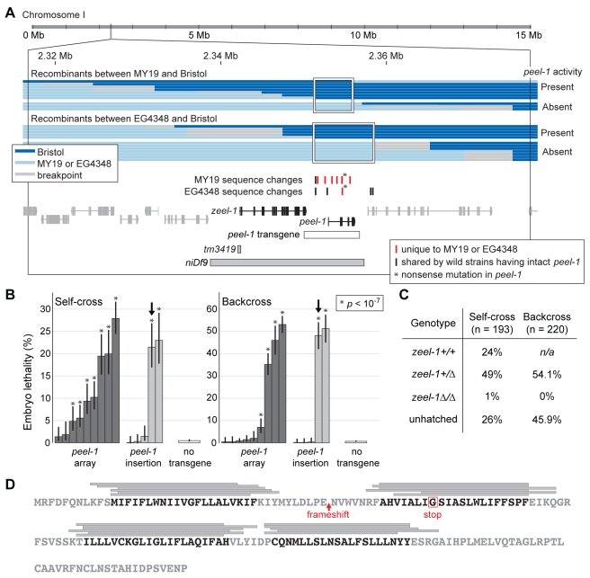
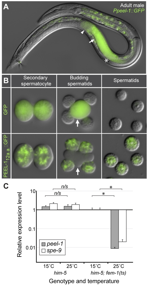
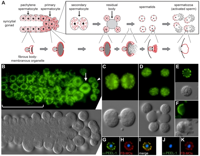
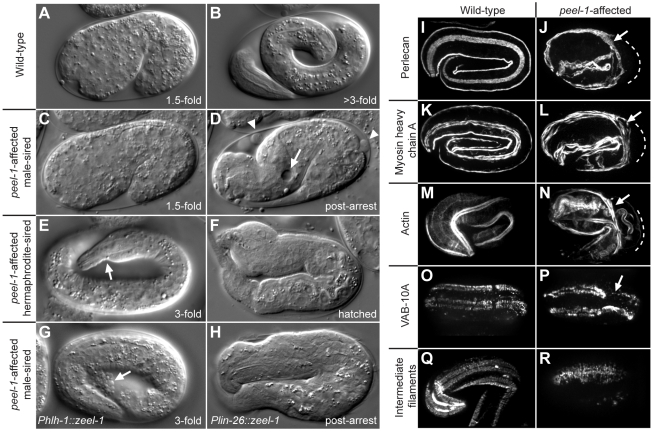
The evolutionary fate of an allele ordinarily depends on its contribution to host fitness. Occasionally, however, genetic elements arise that are able to gain a transmission advantage while simultaneously imposing a fitness cost on their hosts. We previously discovered one such element in C. elegans that gains a transmission advantage through a combination of paternal-effect killing and zygotic self-rescue. Here we demonstrate that this element is composed of a sperm-delivered toxin, peel-1, and an embryo-expressed antidote, zeel-1. peel-1 and zeel-1 are located adjacent to one another in the genome and co-occur in an insertion/deletion polymorphism. peel-1 encodes a novel four-pass transmembrane protein that is expressed in sperm and delivered to the embryo via specialized, sperm-specific vesicles. In the absence of zeel-1, sperm-delivered PEEL-1 causes lethal defects in muscle and epidermal tissue at the 2-fold stage of embryogenesis. zeel-1 is expressed transiently in the embryo and encodes a novel six-pass transmembrane domain fused to a domain with sequence similarity to zyg-11, a substrate-recognition subunit of an E3 ubiquitin ligase. zeel-1 appears to have arisen recently, during an expansion of the zyg-11 family, and the transmembrane domain of zeel-1 is required and partially sufficient for antidote activity. Although PEEL-1 and ZEEL-1 normally function in embryos, these proteins can act at other stages as well. When expressed ectopically in adults, PEEL-1 kills a variety of cell types, and ectopic expression of ZEEL-1 rescues these effects. Our results demonstrate that the tight physical linkage between two novel transmembrane proteins has facilitated their co-evolution into an element capable of promoting its own transmission to the detriment of organisms carrying it.


Conflict of interest statement

The authors have declared that no competing interests exist.

Figures

Figure 1
Figure 1. peel-1 is located adjacent to zeel-1 and encodes a four-pass transmembrane protein.
(A) The large black box outlines the genomic region to which peel-1 was mapped previously . Horizontal bars represent the recombinants used to map the peel-1 mutations in strains MY19 and EG4348. The maximum intervals defined by these mapping experiments are outlined by white boxes. Adjoining breakpoints are excluded because they contain no sequence changes relative to Bristol. Below the recombinants, tick marks indicate all sequence changes in MY19 or EG4348 located within the boxed intervals. Horizontal bars represent the peel-1 transgene, the zeel-1(tm3419) deletion allele, and the deficiency, niDf9. niDf9 is carried by the Hawaii strain and by all other wild isolates lacking the activities of both peel-1 and zeel-1 . (B) The Bristol allele of the peel-1 transgene shown in (A) was tested for its ability to restore peel-1 activity to animals carrying the peel-1 nonsense mutation found in EG4348. To test for peel-1 activity, transgenic animals were crossed to the Hawaii strain, and embryo lethality was scored from self-fertilizing F1 hermaphrodites (self-cross) and F1 males backcrossed to Hawaii hermaphrodites (backcross). Nine independent extra-chromosomal arrays and five independent single-copy genomic insertions were tested. For each array or insertion, 200 to 650 embryos were scored per self-cross or backcross. Ten control replicates were performed in parallel, each including 200 to 400 embryos. The global mean of these replicates is shown by the “no transgene” bars, and lethality for each individual replicate was less than 1.5%. Error bars indicate 95% binomial confidence intervals, calculated using the Agresti-Coull method . * p<10−7, one-tailed binomial test relative to the mean of the control replicates. The observed variability among extra-chromosomal arrays was expected because of germline silencing . The three single-copy insertions showing no peel-1 activity probably represent incomplete insertion events, which are a common outcome of the MosSCI method . Arrows indicate the insertion used for further analysis in (C). (C) To confirm that the lethality observed in (B) was limited to zeel-1(Δ) embryos, an additional self-cross and backcross were performed using the insertion marked in (B). All hatched progeny were genotyped at zeel-1. In both crosses, the genotype frequencies among surviving progeny differed significantly from their Mendelian expectations (χ2 tests, p<10−9). n/a, not applicable. (D) The amino acid sequence of PEEL-1. Grey bars indicate predicted transmembrane helices, as predicted by (from top to bottom): TopPred , Tmpred , TMHMM , SOSUI , PHDhtm , and HMMTOP . Regions predicted by at least four algorithms are highlighted in black. The frameshift in MY19 and the stop codon in EG4348 are indicated in red.
Figure 2
Figure 2. peel-1 is expressed exclusively in sperm and carries an N-terminal sperm localization signal.
(A) Adult male expressing Ppeel-1::GFP. The tube-like gonad begins at the asterisk, extends toward the head, folds over on itself, and then extends toward the tail. The arrow and arrowhead indicate spermatocytes and sperm, respectively. Fluorescence outside the gonad is auto-fluorescence. Nomarski and fluorescence channels are overlaid. (B) Secondary spermatocytes, budding spermatids, and mature spermatids expressing either untagged GFP or GFP tagged with the N-terminal 12 amino acids of PEEL-1 (PEEL-112a.a.::GFP). Arrows indicate residual bodies. Nomarski and fluorescence channels are overlaid. (C) Relative expression levels of peel-1 and spe-9 in him-5(e1490) and him-5(e1490) fem-1(hc17ts) adult males at the permissive (15°C) and restrictive (25°C) temperatures. him-5(e1490) was included to aid in collection of males. Expression levels were calculated relative to the him-5(e1490) fem-1(hc17ts) 15°C sample. Runs were performed in triplicate and standard deviations are shown. * p<10−5, one-tailed Student's t test on the normalized expression levels. n/s, p>0.05.
Figure 3
Figure 3. PEEL-1 localizes to fibrous body-membranous organelles.
(A) Diagram of spermatogenesis and fibrous body-membranous organelle (FB-MO) development, adapted with permission from . FB-MOs develop in pachytene spermatocytes as membrane-bound organelles having a head region separated by a collar-like constriction from a set of membrane folds (lower panel; red). As spermatogenesis proceeds, the membrane folds grow and extend into arm-like protrusions, enveloping bundles of polymerized Major Sperm Protein, referred to as fibrous bodies (hatched region). Coincident with budding of spermatids from the residual body, the membrane folds of FB-MOs retract, and the fibrous bodies depolymerize into the cytoplasm. The FB-free MOs then move to a position just inside the plasma membrane, and upon sperm activation, they fuse with the plasma membrane opposite the pseudopod. (B–F) Nomarski and fluorescence images of spermatocytes and sperm expressing PEEL-1::GFP. Panels in (B) show the proximal arm of a male gonad, oriented with pachytene spermatocytes towards the left (bracketed region). Arrow and arrowhead indicate primary and secondary spermatocytes, respectively. Panels in (C–F) show higher resolution images of the following stages: secondary spermatocyte (C), budding spermatids (D), unactivated spermatid (E), and activated spermatozoan (F). (G–K) Spermatids were dissected from peel-1(+) (G–I) or peel-1(Δ) (J–K) males and stained with anti-PEEL-1 (green) and the FB-MO marker, 1CB4 (red) . Nuclei are stained with DAPI (blue).
Figure 4
Figure 4. peel-1-affected embryos exhibit late-occurring defects in muscle and epidermal tissue.
(A and B) Wild-type embryo at the 1.5-fold stage (A) and just before hatching (B). (C and D) peel-1-affected, male-sired embryo at the 1.5-fold stage (C) and approximately 4 h after the 2-fold arrest (D). Relative to its shape at the 2-fold stage, the embryo in (D) has shortened longitudinally and thickened circumferentially. Thinning of the tail, distention of the excretory cell (arrow), and epidermal leakage (arrowheads) are visible. (E and F) peel-1-affected, hermaphrodite-sired embryos displaying less severe phenotypes than the embryo shown in (D). In (E), the embryo has elongated past the 2-fold stage, but muscle detachment is visible (arrow). In (F), the embryo has hatched but is severely deformed. (G–H) peel-1-affected, male-sired embryos expressing zeel-1 in only muscle (G) or only in epidermis (H). The embryo in (G) has elongated past the 2-fold stage, but epidermal leakage is visible (arrow). The embryo in (H) has arrested paralyzed at the 2-fold stage but has survived to hatching. (I–N) Perlecan, myosin heavy chain A, and F-actin were visualized in wild-type and peel-1-affected, male-sired embryos. In peel-1-affected embryos, muscle detachment is evident at the mid-embryo bend, where muscle fibers (arrows) are displaced inward relative to their proper locations (dashed lines). (O–R) VAB-10A and intermediate filaments were visualized in wild-type and peel-1-affected, male-sired embryos. In peel-1-affected embryos, VAB-10A and intermediate filaments are recruited properly to the four muscle quadrants, but they do not organize into evenly spaced, circumferentially oriented bands. In (P), VAB-10A staining is absent in one dorsal quadrant at the mid-embryo bend (arrow). Images in (O–P) are dorsal views.
Figure 5
Figure 5. The phenotypic effects of sperm-supplied PEEL-1 are dose-dependent.
(A) The proportion of embryos arresting at the 2-fold stage was calculated among peel-1-affected embryos sired by 1- to 3-d-old hermaphrodites. Within each age class, embryos derive from a total of approximately 50 to 150 hermaphrodites. All pair-wise combinations of age classes were compared using χ2 tests. For all pairs, p<10−5. (B) Embryo lethality was scored among peel-1-affected embryos sired by unmated, 1- to 5-d-old hermaphrodites and by partially mated, 3- to 5-d-old hermaphrodites. In the unmated experiment, 91 hermaphrodites were followed from the onset of adulthood, and all embryos laid during the first 5 d of adulthood were scored. The results for 10 randomly selected broods are shown in Figure S4. To generate embryos sired by partially mated animals, 130 hermaphrodites were briefly mated to males following the L4 molt, and embryos were collected during days 3 to 5 from the 35 hermaphrodites that produced a mixture of self- and cross-progeny. Self- and cross-progeny were distinguished by the use of an integrated GFP marker carried by the male, and cross-progeny were excluded from analysis. χ2 tests were used to compare the unmated and partially mated “3–5d” age classes, as well as all pair-wise combinations of age classes within the unmated experiment. n/s, p>0.05. * and all unlabeled pairs within the unmated experiment, p<10−5. (C) Embryo lethality was scored among peel-1-affected embryos derived from crosses between (i) 1-d-old males and hermaphrodites, (ii) 3-d-old males and hermaphrodites, and (iii) 5-d-old hermaphrodites that had been removed from males on day 2 in order to allow male sperm to age for 3 d within the reproductive tract of the hermaphrodite. In each cross, embryos derive from a total of 12 to 16 hermaphrodites. (D) Spindle plots showing the onset of epidermal leakage in peel-1-affected embryos sired by hermaphrodites carrying one or two copies of peel-1(+), or by males carrying one, two, or three copies of peel-1(+). A third copy of peel-1 was added using the single-copy insertion of the peel-1 transgene marked in Figure 1B. The width of each bar reflects the proportion of embryos initiating leakage in each time interval. All pair-wise combinations of spindle plots were compared using Mann-Whitney U tests on the raw distributions of leakage times. n/s, p>0.05. For all other pairs, p<10−5.
Figure 6
Figure 6. zeel-1 is transiently expressed during embryogenesis and localizes to cell membranes.
(A) zeel-1 mRNAs were quantified in wild-type embryos via single-molecule fluorescence in situ hybridization . Embryos were staged by counting nuclei manually (1- to 40-cell embryos), counting nuclei using image analysis software (41- to 200-cell embryos), or classifying embryos as end of gastrulation (∼250 cells), comma, 2-fold, or ≥3-fold. Inset shows a magnification of the boxed area. Each circle represents an independent embryo. n = 130. (B–C) Embryos expressing ZEEL-1::GFP. Panel in (B) shows a dorsal view during intercalation of epidermal cells. Arrow indicates an epidermal cell membrane. Panel in (C) shows a lateral cross-section of a 1.5-fold embryo. The apical face of the pharynx (arrow) and the intestine (arrowhead) are indicated.
Figure 7
Figure 7. The transmembrane domain of zeel-1 is evolutionarily novel and partially sufficient for function.
(A) Maximum likelihood phylogeny of the protein sequences of all zyg-11 homologs in C. elegans. Genes containing predicted transmembrane domains are highlighted in black. The transmembrane domains of these genes were excluded prior to analysis. Genes located in the tandem array are highlighted by a shaded grey rectangle. Scale bar indicates amino acid substitutions per site. Values on branches indicate percent bootstrap support. The asterisk indicates that the reference sequence of Y71A12B.18 contains a single frame-shift, corrected prior to analysis. (B) Four zeel-1-derived transgenes were introduced into a strain carrying the zeel-1 deficiency, niDf9, and tested for their ability to rescue peel-1-affected embryos. To test for rescue, transgenic animals were crossed to the Bristol strain, and lethality was scored among embryos derived from self-fertilizing F1 hermaphrodites (self-cross) and F1 males backcrossed to hermaphrodites of the original transgenic line (backcross). For each type of transgene, 4 to 13 independent extra-chromosomal arrays were tested. For each array, 100 to 600 embryos were scored per self-cross or backcross. Ten control replicates were performed in parallel, each including 100 to 400 embryos (“no transgene” bars). Among arrays or control replicates, lethality scores were averaged to obtain global means and standard deviations. Each transgene was tested for a reduction in lethality compared to the control replicates (Student's t tests, p values indicated by shading). Additionally, the two rescuing transgenes (ZEEL-1TM and ZEEL-1::GFP) were tested for significant differences relative to one another (Student's t tests; * p<0.005; n/s, p>0.05). For the rescuing transgenes, lethality was not reduced to zero because extra-chromosomal arrays are not transmitted to all progeny. (C) Hatch rates were compared between peel-1-affected embryos that we confirmed to have inherited either ZEEL-1TM or ZEEL-1::GFP2 tests, p values shown). Separate comparisons were performed for male- and hermaphrodite-sired embryos. Unless otherwise specified, all hatched progeny appeared morphologically normal. Inheritance of the transgenes was determined by expression of the co-injection marker, Pmyo-2::RFP. Sibling embryos not inheriting the transgene were used as internal negative controls. The hatch rates of these controls were 2% (n = 601−653) among hermaphrodite-sired embryos and 0% (n = 341−933) among male-sired embryos.
Figure 8
Figure 8. Ectopic expression of peel-1 and zeel-1 replicates peel-1-mediated toxicity and zeel-1-mediated rescue.
(A) Survival curves for heat-shocked adult animals carrying extra-chromosomal arrays or single-copy genomic insertions of Phsp-16.2::peel-1 or Phsp-16.41::peel-1. Time zero indicates the start of a 1-hour heat-shock at 34°C. Curves represent one array and one insertion of Phsp-16.2::peel-1 and two independent arrays and seven independent insertions of Phsp-16.41::peel-1. Log-rank tests were used to compare (i) insertions versus arrays and (ii) Phsp-16.2::peel-1 versus Phsp-16.41::peel-1. Data for independent arrays or insertions of Phsp-16.41::peel-1 were combined prior to analysis. * p<10−16. n/s, p>0.05. n = 40–70 animals per curve. (B) One array and one insertion of Phsp-16.41::peel-1 were chosen from (A) to be tested against five independent arrays and one single-copy insertion of Phsp-16.41::zeel-1. Animals carrying both types of transgenes were heat-shocked as in (A). Assays were truncated at 10 h post-heat-shock. Log-rank tests were used to compare each assay to the corresponding assay of Phsp-16.41::peel-1 alone from (A) (p values shown). n = 45−180 animals per curve. (C) The following classes of embryos, aged 3 to 7.5 h post-four-cell stage, were heat-shocked for 20 min at 34°C: (i) zeel-1(Δ) embryos carrying a Phsp-16.41::peel-1 array (pale green bars), (ii) zeel-1(Δ) and zeel-1(+) embryos carrying a Phsp-16.41::peel-1 insertion (black and grey bars, respectively), and (iii) peel-1-affected, male-sired embryos carrying either an array or an insertion of Phsp-16.41::zeel-1 (yellow and red bars, respectively). For each genotypic class, the proportion of embryos arresting at the 2-fold stage relative to all embryos that elongated to the 2-fold stage is plotted. Differences between the black and grey bars are not significant (p>0.05, χ2 tests for each age class). See Figure S8 for the full dataset. (D–E) Vulva regions of animals carrying an array of Pexp-3::peel-1 and an integrated copy of Pmyo-3::GFP, a marker of the egg-laying muscles. Somatic inheritance of the Pexp-3::peel-1 array was followed by co-injection markers, Prab-3::mCherry and Pmyo-3::mCherry, which express in neurons and the egg-laying and body-wall muscles, respectively. In (D), the egg-laying muscles (arrows) are morphologically normal and have not inherited the Pexp-3::peel-1 array, as indicated by absence of mCherry expression. In (E), the left-hand egg-laying muscle is severely atrophied (arrow) and the right-hand egg-laying muscle is absent. The atrophied muscle cell expresses mCherry, indicating this cell has inherited the array of Pexp-3::peel-1. In both images, mCherry expression is also visible in body-wall muscles and neighboring neurons. (F) Dead GABA neuron (arrow) in an animal carrying an array of Punc-47::peel-1. The surrounding tissue, including a neighboring, non-GABA neuron (arrowhead), is morphologically normal.

References

    1. Hurst G. D. D, Werren J. H. The role of selfish genetic elements in eukaryotic evolution. Nature Reviews Genetics. 2001;2:597–606. - PubMed
    1. Dawe R. K, Hiatt E. N. Plant neocentromeres: fast, focused, and driven. Chromosome Res. 2004;12:655–669. - PubMed
    1. Fishman L, Willis J. H. A novel meiotic drive locus almost completely distorts segregation in Mimulus (monkeyflower) hybrids. Genetics. 2005;169:347–353. - PMC - PubMed
    1. Pardo-Manuel de Villena F, Sapienza C. Nonrandom segregation during meiosis: the unfairness of females. Mamm Genome. 2001;12:331–339. - PubMed
    1. Jaenike J. Sex chromosome meiotic drive. Annual Review of Ecology and Systematics. 2001;32:25–49.

Publication types

MeSH terms