. 2011 Jul;7(7):e1002088.
doi: 10.1371/journal.pcbi.1002088. Epub 2011 Jul 21.

The Eps8/IRSp53/VASP network differentially controls actin capping and bundling in filopodia formation

Affiliations

The Eps8/IRSp53/VASP network differentially controls actin capping and bundling in filopodia formation

Federico Vaggi et al. PLoS Comput Biol. 2011 Jul.

Abstract

There is a body of literature that describes the geometry and the physics of filopodia using either stochastic models or partial differential equations and elasticity and coarse-grained theory. Comparatively, there is a paucity of models focusing on the regulation of the network of proteins that control the formation of different actin structures. Using a combination of in-vivo and in-vitro experiments together with a system of ordinary differential equations, we focused on a small number of well-characterized, interacting molecules involved in actin-dependent filopodia formation: the actin remodeler Eps8, whose capping and bundling activities are a function of its ligands, Abi-1 and IRSp53, respectively; VASP and Capping Protein (CP), which exert antagonistic functions in controlling filament elongation. The model emphasizes the essential role of complexes that contain the membrane deforming protein IRSp53, in the process of filopodia initiation. This model accurately accounted for all observations, including a seemingly paradoxical result whereby genetic removal of Eps8 reduced filopodia in HeLa, but increased them in hippocampal neurons, and generated quantitative predictions, which were experimentally verified. The model further permitted us to explain how filopodia are generated in different cellular contexts, depending on the dynamic interaction established by Eps8, IRSp53 and VASP with actin filaments, thus revealing an unexpected plasticity of the signaling network that governs the multifunctional activities of its components in the formation of filopodia.


Conflict of interest statement

The authors have declared that no competing interests exist.

Figures

Figure 1
Figure 1. Eps8 and IRSp53 effector network.
Network showing the main interactors of Eps8 and IRSp53 involved in the regulation of filopodia formation. Black filled dots indicate substrates of a reversible binding reaction, whose product is pointed by an arrow. Turnover of filamentous actin (reaction (9)) is the only irreversible reaction depicted in the diagram. In the network, we identify two different modules, a capping module, which includes the binding reactions between cappers and barbed ends, and a bundling module, which includes the binding reactions between bundlers and filamentous actin. CP represents Capping Protein, cyan circles are polymerized monomers of actin; the red circle marked Ga is G-actin. B and P mark the barbed and pointed ends, respectively, of a filament of actin. Reaction numbers and the shortened names in parentheses under the icons allow an easy interpreation of the equations of the model (Table 1 in Text S1).
Figure 2
Figure 2. VASP synergizes with IRSp53 in bundling actin filaments and competes with Eps8 for IRSp53 binding.
a. Isolated VASP and Eps8 bundle actin filaments with low efficiency, which is enhanced by their association with IRSp53. The bundling efficiency was determined by measuring the number of bundles/field obtained in fluorescence microscopy-based F-actin-bundling assays as described and shown in Fig. S1A–B. At least 10 fields per experiment performed in triplicates were scored. Data are the mean ± s.e.d. b. Measurement of IRSp53 and VASP interaction. Equal amounts (10 pmoles) of His-IRSp53, GST-IRSp53-SH3 or BSA were spotted onto nitrocellulose and incubated with increasing concentrations of purified VASP. The nitrocellulose filter was then subjected to WB analysis using anti-VASP antibody (Ab). The fraction of VASP bound was plotted against the concentrations of total VASP. An apparent dissociation constant was calculated using standard procedure as described in . c. The proline rich region of Eps8 (PPP) competes with VASP for binding to IRSp53. Equal amounts (10 pmoles) of His-IRSp53 spotted onto nitrocellulose and incubated with purified 100 nM VASP or BSA as control, in the absence or the presence of increasing amounts of the proline-rich region of Eps8 (GST-PPP) or GST. The filters were immunoblotted with the indicated abs. d. VASP forms a complex with IRSp53 in-vivo. Lysates (1 mg) of HeLa cells were immunoprecipitated with anti-VASP or with control abs. Lysates (20 µg) and immunoprecipitates (IP) were immunoblotted with the indicated abs. The bottom panel is a longer exposure to visualize endogenous levels of VASP.
Figure 3
Figure 3. Average concentrations of filopodia initiator correlates with the probability of forming filopodia.
a. Distributions of the concentration of filopodia initiators (FI) in cell populations with different mean values (μ) and identical standard deviationσ, computed as formula image
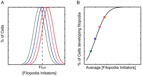
. The concentration of FI required for initiating the positive feedback loop (FIcrit) is shown as a dotted line. As μ increases (different colored curves) the fraction of cells with FI>FIcrit increases. b. Fraction of cells in a population with FI>FIcrit as a function of the average FI concentration μ. Different color squares represent the fraction of cells for the different Gaussians shown in A. To calculate the amount of cells with FI>FIcrit, we simply integrate the Gaussian from FIcrit to infinite, formula image.
Figure 4
Figure 4. Eps8 plays a major role as a bundler, and not as a capper, in HeLa cells.
a. Change in RFI and FII in the various genetic backgrounds. Empty rectangles represent experimental results (see Table 3 in Text S1), filled rectangles simulations of equations in Table 1 in Text S1 and parameters in Table 2 and Table 3 in Text S1. b. Complexes formed in HeLa cells by Abi1, Eps8, IRSp53, and VASP in different genetic backgrounds, plotted as percentage of total protein concentration in the wild type. Simulations performed as in a. c. Removal of Eps8 from HeLa cells does not significantly increase the amount of VASP bound to IRSp53. Lysates (1 mg) of HeLa control cells treated with a scrambled oligo [WT (scr)] or interfered for Eps8 (Eps8 K.d.) were immunoprecipitated with VASP or control abs. Lysates (40 µg) and immunoprecipitates (IPs) were immunoblotted with the indicated abs. IgG are also indicated.
Figure 5
Figure 5. In Neurons Eps8 prevalently acts as a capper.
a. Change in RFI and FII (i.e., Eps8:IRSp53:Fa and VASP:IRSp53:Fa normalized with respect to their concentrations in wild type cells) in the various genetic backgrounds. Empty rectangles represent experimental results (see Table 3 in Text S1), filled rectangles reproduce simulations of equations in Table 1 in Text S1 and parameters in Table 2 and Table 3 in Text S1. b. Complexes formed in HeLa cells by Abi1, Eps8, IRSp53, and VASP in the different genetic backgrounds plotted as percentage of total protein concentration in the wild type. Simulations performed as in a. c. Removal of Eps8 from neurons significantly increases the amount of VASP bound to IRSp53. Cortex and hippocampus lysates (1 mg) derived from Eps8 WT or KO mice were immunoprecipitated with anti-IRSp53 or anti Flag as control. Lysates (20 µg) and immunoprecipitates (IPs) were immunoblotted with the indicated abs.
Figure 6
Figure 6. Eps8:IRSp53 induces filopodia formation in VASP-deficient MVD7 cells after RNAi-mediated removal of CP.
a. RNAi-mediated downregulation of CP in MVD7 cells over-expressing IRSp53 increases filopodia formation. Control (WT scr) or CP (CP KD) RNAi-treated MVD7 cells transfected with Flag–IRSp53 were fixed and stained with rhodamine–phalloidine or anti-flag to detect F-actin (red) or IRSp53 (blue), respectively. Right panels, magnifications corresponding to the white dashed squares of the pictures on the left (the different channels are indicated). DI are digitalized images obtained with Adobe Photoshop filters starting from the actin channel to highlight cells protrusions . Bar is 10 µm (4 µm for the magnifications). b. The expression of endogenous CP in cells interfered for CP (CP KD) or treated with scrambled oligo (WT scr) was analyzed by immunoblotting with the indicated abs. CP reduction (85%) was determined using the software ImageJ, by analyzing the intensity of the signals for CP in control cells (WT scr) or cells interfered for CP (CP KD), normalizing over vinculin signal. c. Change in RFI and FII (i.e., Eps8:IRSp53:Fa normalized by its wild type value, see main text) in WT and CP knockout MVD7 cells. Empty rectangles represent experimental results, filled rectangles simulations of equations in Table 1 in Text S1 and parameters in Table 2 and Table 3 in Text S1. d. Complexes formed in HeLa cells by Eps8, IRSp53 and Abi1 in CP Kd and WT plotted as percentage of total protein concentration in the wild type. Simulations as in c.

References

    1. Mattila PK, Lappalainen P. Filopodia: molecular architecture and cellular functions. Nat Rev Mol Cell Biol. 2008;9:446–454. - PubMed
    1. Mogilner A, Rubinstein B. The physics of filopodial protrusion. Biophys J. 2005;89:782–795. - PMC - PubMed
    1. Lebrand C, Dent EW, Strasser GA, Lanier LM, Krause M, et al. Critical role of Ena/VASP proteins for filopodia formation in neurons and in function downstream of netrin-1. Neuron. 2004;42:37–49. - PubMed
    1. Breitsprecher D, Kiesewetter AK, Linkner J, Urbanke C, Resch GP, et al. Clustering of VASP actively drives processive, WH2 domain-mediated actin filament elongation. EMBO J. 2008;27:2943–2954. - PMC - PubMed
    1. Pasic L, Kotova T, Schafer DA. Ena/VASP proteins capture actin filament barbed ends. J Biol Chem. 2008;283:9814–9819. - PMC - PubMed

Publication types

MeSH terms