. 2011 Jul;7(7):e1002098.
doi: 10.1371/journal.ppat.1002098. Epub 2011 Jul 21.

A role for the chemokine RANTES in regulating CD8 T cell responses during chronic viral infection

Affiliations

A role for the chemokine RANTES in regulating CD8 T cell responses during chronic viral infection

Alison Crawford et al. PLoS Pathog. 2011 Jul.

Abstract

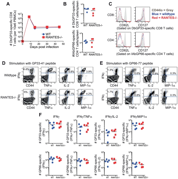
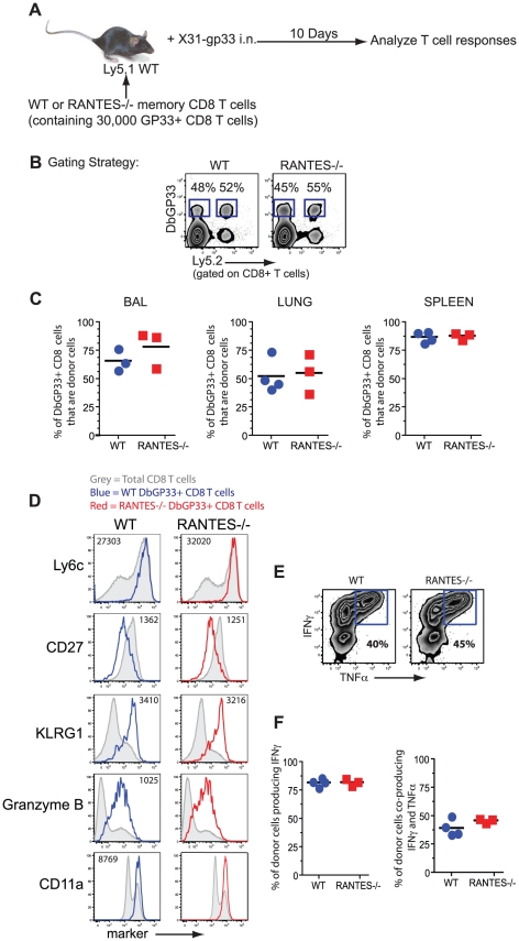
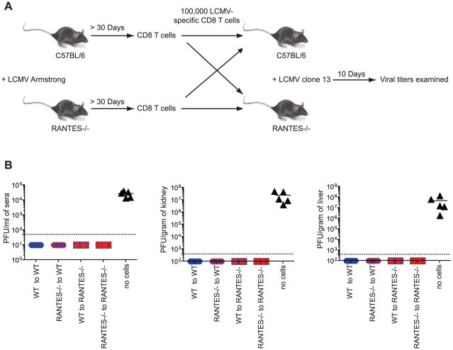
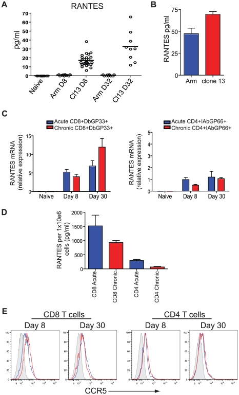
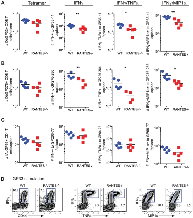
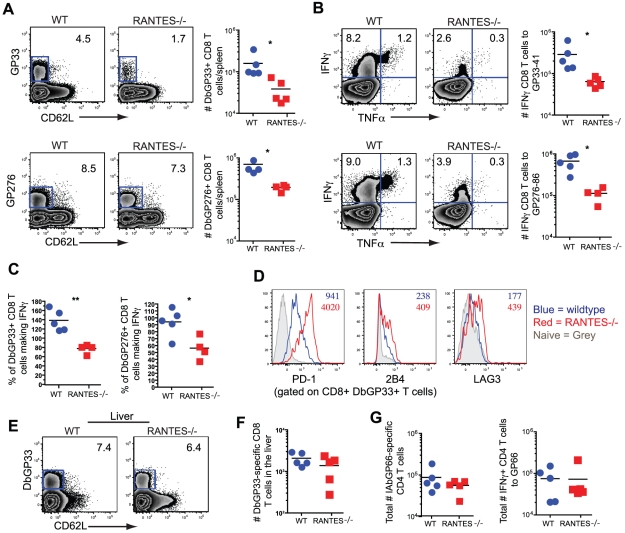
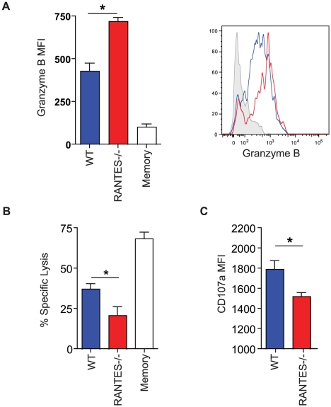
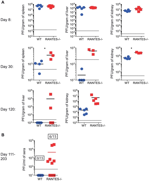
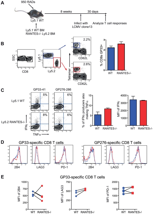
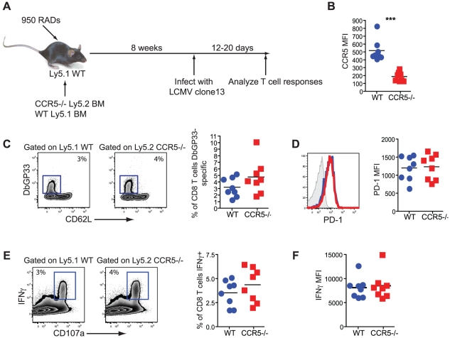
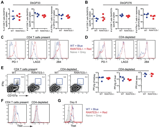
RANTES (CCL5) is a chemokine expressed by many hematopoietic and non-hematopoietic cell types that plays an important role in homing and migration of effector and memory T cells during acute infections. The RANTES receptor, CCR5, is a major target of anti-HIV drugs based on blocking viral entry. However, defects in RANTES or RANTES receptors including CCR5 can compromise immunity to acute infections in animal models and lead to more severe disease in humans infected with west Nile virus (WNV). In contrast, the role of the RANTES pathway in regulating T cell responses and immunity during chronic infection remains unclear. In this study, we demonstrate a crucial role for RANTES in the control of systemic chronic LCMV infection. In RANTES⁻/⁻ mice, virus-specific CD8 T cells had poor cytokine production. These RANTES⁻/⁻ CD8 T cells also expressed higher amounts of inhibitory receptors consistent with more severe exhaustion. Moreover, the cytotoxic ability of CD8 T cells from RANTES⁻/⁻ mice was reduced. Consequently, viral load was higher in the absence of RANTES. The dysfunction of T cells in the absence of RANTES was as severe as CD8 T cell responses generated in the absence of CD4 T cell help. Our results demonstrate an important role for RANTES in sustaining CD8 T cell responses during a systemic chronic viral infection.


Conflict of interest statement

The authors have declared that no competing interests exist.

Figures

Figure 1
Figure 1. The absence of RANTES does not affect T cell responses to acute LCMV infection.
WT and RANTES−/− mice were infected with LCMV Armstrong. Mice were bled on days 8, 15, 30 and 45 p.i. and T cell responses examined. (A) The frequency of DbGP33-specific CD8 T cells in the blood in WT and RANTES−/− mice was determined. Splenocytes from WT and RANTES−/− mice were examined 52 days p.i. (B–F). Total numbers of DbGP33-specific CD8 and IAbGP66-specific CD4 T cells in the spleen at the memory phase of the response were determined using tetramers (B). CD62L and CD127 expression was examined on LCMV-specific memory CD4 and CD8 T cells from both WT and RANTES−/− mice (C). Splenocytes from WT and RANTES−/− mice were stimulated with GP33 and GP66 peptides to measure cytokine responses from CD8 and CD4 T cells, respectively (D–F). The cytokines IFNγ, TNFα, IL-2 and MIP-1α were measured and representative FACs plots are shown. Graphs show total numbers per spleen. Data are representative of 2 independent experiments with at least 4 mice per group in each experiment.
Figure 2
Figure 2. WT and RANTES−/− mice mount equivalent secondary responses.
(A) WT and RANTES−/− mice were infected with LCMV Armstrong. After ∼50 days, CD8 T cells were purified from the mice and equal numbers of WT or RANTES−/− DbGP33-specific CD8 T cells were transferred into congenically marked Ly5.1 mice. The mice were then infected the following day with X31-gp33 i.n. Ten days later, the number of donor (Ly5.2+) virus-specific CD8 T cells was enumerated by tetramer staining. Gating strategy to identify donor responses (B). WT and RANTES−/− memory T cells were measured in the BAL (bronchoalveolar lavage), lung and spleen of recipient mice (C). WT and RANTES−/− DbGP33-specific CD8 T cells were examined in the spleen and stained for Ly6c, CD27, KLRG1, granzyme B and CD11a (D). Production of IFNγ or TNFα by the adoptively transferred cells was measured in the spleen by peptide stimulation and ICS (E and F). Data are representative of 2 independent experiments each with 3 mice per group.
Figure 3
Figure 3. Memory CD8 T cells do not need RANTES to protect from chronic LCMV infection.
(A) WT and RANTES−/− mice were infected with LCMV Armstrong. Over 30 days later, CD8 T cells were purified from the mice and 100,000 WT or RANTES−/− DbGP33-specific CD8 T cells were adoptively transferred into WT or RANTES−/− mice (A). The mice were then infected the following day with LCMV clone 13 i.v. Nine days later, viral titers were measured in the spleen, kidney and sera of these mice (B). The results of two independent experiments shown in the same graph.
Figure 4
Figure 4. Higher concentrations of RANTES protein are present in the serum of mice infected with LCMV clone 13 compared to LCMV Armstrong and naïve mice.
(A) C57Bl/6 mice were infected with LCMV Armstrong or LCMV clone 13 and the sera examined 8 and 32 days later for RANTES protein using luminex. (B) C57Bl/6 mice were infected with LCMV Armstrong or LCMV clone 13 and the sera examined 6 days later for RANTES protein by ELISA. A total of three mice each were infected. Data are representative of 2 independent experiments. (C) Naïve CD8 T cells and DbGP33-specific CD8 T cells from day 8 and day 30 p.i. with LCMV Armstrong or clone 13 were examined for expression of RANTES mRNA by RT-PCR. (D) Mice were infected with LCMV Armstrong or clone 13 and CD44hi CD4 and CD8 T cells sorted on day 8 p.i. Sorted CD8 T cells were incubated with PMA/ionomycin for 5 hours and the supernatants examined for RANTES protein. (E) C57Bl/6 mice were infected with LCMV Armstrong or LCMV clone 13 and 8 and 30 days p.i. DbGP33-specific CD8 T cells were examined for expression of CCR5 (grey = naïve, blue = LCMV Armstrong, red = LCMV clone 13.
Figure 5
Figure 5. The primary CD8 T cell cytokine response is diminished in the absence of RANTES at one week after clone 13 infection.
WT and RANTES−/− mice were infected with LCMV clone 13 and T cell responses examined eight days later. The total number of LCMV-specific CD8 and CD4 T cells was measured by tetramer staining as well as peptide stimulation and ICS to detect cytokine production. Expression of IFNγ, TNFα and MIP1α was examined (A–D). Graphs show total numbers per spleen. Representative plots of IFNγ, TNFα and MIP-1α expression (D). Data are representative of 3 independent experiments with five mice per group.
Figure 6
Figure 6. CD8 T cell responses are significantly reduced in RANTES−/−mice one month p.i.
The total numbers of LCMV-specific CD8 T cells were examined in WT and RANTES−/− mice 30 days p.i. with LCMV clone 13. Representative FACs plots are shown on the left (A). The ability to produce IFNγ was measured by peptide stimulation and ICS (B). The percentage of LCMV tetramer +ve cells able to make IFNγ was calculated from the total number of tetramer +ve CD8 T cells and total number of IFNγ-producing T cells in response to the same peptide (C). DbGP33-specific CD8 T cells from WT and RANTES−/− mice were stained for PD-1, 2B4 and LAG3. Representative plots are shown. Numbers represent the MFI. Grey = naïve, blue = DbGP33-specific CD8 T cells from acute infection, red = DbGP33-specific CD8 T cells from chronic infection (D). DbGP33-specific CD8 T cells were examined in the liver. Representative plots with numbers indicating the percentage of CD8 T cells that were DbGP33-specific (E). Total numbers of DbGP33-specific CD8 T cells in the liver were determined by tetramer staining. (F). The total number of IAbGP66-speciifc CD4 T cells were determined by tetramer staining as well as peptide stimulation and ICS (G). Data are representative of three independent experiments each with at least four mice per group.
Figure 7
Figure 7. The cytotoxic ability of virus-specific CD8 T cells was decreased in the absence of RANTES.
WT and RANTES−/− mice were infected with LCMV clone 13 and 30 days later. LCMV-specific CD8 T cells were examined for expression of granzyme B (A). Results are shown graphically (left) and a representative histogram is shown. Grey = Naïve CD8 T cells, Blue = DbGP33-specific CD8 T cells from acute infection, red = DbGP33-specific CD8 T cells from chronic infection. Equal number of GP33-specific CD8 T cells were examined for their ability to kill CFSE-labeled target cells (B). Surface CD107a was measured during a 5 hour stimulation (C).
Figure 8
Figure 8. Higher viral loads later during chronic LCMV infection in RANTES−/− versus WT mice.
WT and RANTES−/− mice were infected with LCMV clone 13 and viral titers were determined by plaque assay from tissues at 8, 30 and 102 days p.i. (A). Sera was also examined on days 111–203 p.i. The graph shows the result of three independent experiments shown together (B). Ratios show the number of mice that were viremic in the WT and RANTES−/− groups out of a total of 15 mice.
Figure 9
Figure 9. CD8 T cells do not need to produce RANTES themselves.
Mixed bone-marrow chimeras were made where ∼50% of the cells were WT Ly5.1+ and ∼50% RANTES−/− Ly5.2+ (A). Upon reconstitution, mice were infected with LCMV clone 13 and the CD8 T cell responses analyzed. RANTES−/− T cells were identified by staining with Ly5.2 (B). Both the RANTES−/− and WT CD8 T cells were examined for their ability to produce IFNγ and TNFα (C). Representative FACs plots are shown (left) as well as a bar graph summarizing the percentage of IFNγ-producers able to make TNFα and a graph of the MFI of IFNγ. Both WT and RANTES−/− T cells were stained for 2B4, LAG3 and PD-1 with (D) showing representative plots of staining. Graphs plot the MFI of 2B4, LAG3 and PD-1 (E).
Figure 10
Figure 10. CD8 T cells do not need to bind RANTES directly.
Mixed bone-marrow chimeras were made where ∼50% of the cells were Ly5.1+ WT and ∼50% Ly5.2+ CCR5−/−. Upon reconstitution, mice were infected with LCMV clone 13 and the CD8 T cell responses analyzed 12–20 days later (A). The WT and CCR5−/− IAbGP33-specific CD8 T cells were stained for CCR5 (B). The percentage of WT and CCR5−/− CD8 T cells that were GP33-specific were determined by DbGP33 tetramer (C). Representative staining of PD-1 on DbGP33-specific CD8 T cells and a graph of the MFI is shown (D). IFNγ production was measured in response to GP33-44 peptide stimulation (E). Representative FACs plots are shown (left) as well as graphs showing the percentage of the WT or CCR5−/− CD8 T cells producing IFNγ and the MFI of IFNγ (F). Data are representative of 2 independent experiments each with at least 5 mice per group.
Figure 11
Figure 11. CD4-depletion reduces Tbet and IFNγ production in WT mice similar to levels seen in RANTES−/− mice.
A cohort of WT and RANTES−/− mice were depleted of CD4 T cells with GK1.5 antibody on the day prior to infection with LCMV clone 13. T cell responses were examined 35 days later. (A and B) Percentages and total numbers of LCMV-specific CD8 T cells were determined in WT and RANTES−/− mice depleted of CD4 T cells for both DbGP33-specific and DbGP276-specific CD8 T cells using tetramer. (C) DbGP33 and DbGP276-specific CD8 T cells were examined for expression of PD1, LAG3 and 2B4. Filled grey represents naïve CD8 T cells, blue = DbGP33-specific CD8 T cells from WT mice, red = DbGP33-specific CD8 T cells from RANTES−/− mice. (D) Representative plots of CD107a expression and IFNγ production by DbGP33-specific CD8 T cells. Numbers represent the percent of CD107a+ cells that are also making IFNγ in response to stimulation with GP33-44 peptide. Viral titers were determined by plaque assays in CD4-depleted WT and RANTES−/− mice (E). Tbet expression was determined in DbGP33-specific CD8 T cells by flow cytometry in WT and RANTES−/− mice containing CD4 T cells as well as those depleted of T cells prior to infection (F). Tbet expression was already slightly reduced by 8 days p.i. (G).

References

    1. Sallusto F, Mackay CR, Lanzavecchia A. The role of chemokine receptors in primary, effector, and memory immune responses. Annu Rev Immunol. 2000;18:593–620. - PubMed
    1. Esche C, Stellato C, Beck LA. Chemokines: key players in innate and adaptive immunity. J Invest Dermatol. 2005;125:615–628. - PubMed
    1. Loetscher P, Seitz M, Clark-Lewis I, Baggiolini M, Moser B. Activation of NK cells by CC chemokines. Chemotaxis, Ca2+ mobilization, and enzyme release. J Immunol. 1996;156:322–327. - PubMed
    1. Schall TJ, Bacon K, Toy KJ, Goeddel DV. Selective attraction of monocytes and T lymphocytes of the memory phenotype by cytokine RANTES. Nature. 1990;347:669–671. - PubMed
    1. Rot A, Krieger M, Brunner T, Bischoff SC, Schall TJ, et al. RANTES and macrophage inflammatory protein 1 alpha induce the migration and activation of normal human eosinophil granulocytes. J Exp Med. 1992;176:1489–1495. - PMC - PubMed

Publication types

MeSH terms