. 2011 Nov;130(2):663-79.
doi: 10.1007/s10549-011-1690-0. Epub 2011 Aug 4.

Identification of the receptor tyrosine kinase AXL in breast cancer as a target for the human miR-34a microRNA

Affiliations

Identification of the receptor tyrosine kinase AXL in breast cancer as a target for the human miR-34a microRNA

Mark Mackiewicz et al. Breast Cancer Res Treat. 2011 Nov.

Abstract

The identification of molecular features that contribute to the progression of breast cancer can provide valuable insight into the pathogenesis of this disease. Deregulated microRNA expression represents one type of molecular event that has been associated with many different human cancers. In order to identify a miRNA/mRNA regulatory interaction that is biologically relevant to the triple-negative breast cancer genotype/phenotype, we initially conducted a miRNA profiling experiment to detect differentially expressed miRNAs in cell line models representing triple-negative (MDA-MB-231), ER(+) (MCF7), and HER-2 overexpressed (SK-BR-3) histotypes. We identified human miR-34a expression as being >3-fold down (from its median expression value across all cell lines) in MDA-MB-231 cells, and identified AXL as a putative mRNA target using multiple miRNA/target prediction algorithms. The miR-34a/AXL interaction was functionally characterized through ectopic overexpression experiments with a miR-34a mimic in two independent triple-negative breast cancer cell lines. In reporter assays, miR-34a binds to its putative target site within the AXL 3'UTR to inhibit luciferase expression. We also observed degradation of AXL mRNA and decreased AXL protein levels, as well as cell signaling effects on AKT phosphorylation and phenotypic effects on cell migration. Finally, we present an inverse correlative trend in miR-34a and AXL expression for both cell line and patient tumor samples.


Conflict of interest statement

Conflict of interest: No potential conflicts of interest are disclosed.

Figures

Fig. 1
Fig. 1
miR-34a expression and targeting in triple-negative breast cancer cell lines. a Heat map of the most differentially expressed miRNAs in MDA-MB-231, MCF7, and SK-BR-3 breast cancer cell lines compared to MCF10A cells. Each cell line comparison is represented by four arrays (biological replicates). Hierarchical clustering of samples and genes are shown. Only those human miRNAs with at least a threefold change in expression from its median value across all arrays in at least 33% of the arrays are shown. Red miRNAs that are overexpressed relative to the reference, green miRNAs that are underexpressed relative to the reference. b Gene structure showing the location of the predicted binding site for miR-34a within the AXL 3′UTR, and the structure of the miRNA-target interaction as predicted by the MiRanda program. Seed sequence-binding region is boxed in gray. CDS coding sequence. c Northern blot showing the expression of miR-34a precursor and mature RNAs across 10 different cell lines. Breast cell lines: MCF10A, MCF7, MDA-MB-231, SK-BR-3, Hs578T, T47D, and BT549; renal cell line: SN12C; and colorectal cell lines: SW480 and SW620.U6RNA was probedasaloading control. Asterisks denote triple-negative breast cancer cell lines. High AXL protein expression (see Suppl. Fig. S2) is inversely correlated with low miR-34a expression in these cell lines. d qRT-PCR analysis of mature miR-34a expression in cancer cell lines relative to the reference MCF10A. Error bars represent the range in fold difference. Asterisks denote triple-negative breast cancer cell lines. The results in c and d corroborate with one another, as well as to the relative expression levels observed for miR-34a in those cell lines that were tested on the microarray
Fig. 2
Fig. 2
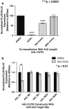
miR-34a targets the 3′UTR of AXL and affects expression of a luciferase reporter construct. Full-length AXL 3′UTR (a) or a 48 bp fragment containing the putative miR-34a target site (b) were cloned into luciferase reporter expression vectors and co-transfected into MDA-MB-231 cells with siNEG siRNA (negative control) siAXL, or mimics for miR-34a and miR-200c. For each construct, normalized firefly luciferase signal for each transfection is presented relative to the siNEG transfected sample. For the full-length 3′UTR of AXL, miR-34a, but not miR-200c, affects luciferase expression; mutations in the seed sequence-binding region for miR-34a nearly abolishes the effect (b). Bars represent the average signal ± SEM
Fig. 3
Fig. 3
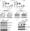
Ectopic miR-34a expression decreases AXL mRNA and protein levels with a downstream effect on AKT phosphorylation.a MDA-MB-231 or Hs578T cells transfected with either siAXL or a miR-34a mimic results in a significant decrease in AXL mRNA levels within these cells. AXL mRNA levels were normalized to PPIB for each transfected sample (data point). Horizontal bar represents the average signal ± SEM. b MDA-MB-231 or Hs578T cells transfected with either siAXL or a miR-34a mimic also results in a significant decrease in AXL protein levels. The receptor tyrosine kinase MET, which was previously shown to be a target for miR-34a, was also affected at the protein level but the results are not as dramatic as those seen with AXL. Values under each band represent the β-Actin normalized intensity of the band relative to that of the siNEG transfected sample. c Decreases in AXL protein levels by either siAXL or the miR-34a mimic affects AKT phosphorylation (at Thr308 and Ser473) in MDA-MB-231 cells with no decrease in total AKT levels. d The effects of siAXL and miR-34a on phospho-AKT (Thr308) are rescued with the co-transfection of an AXL expression vector that lacks the 3′UTR for targeting by these molecules. Western blots are representative of at least duplicate independent experiments
Fig. 4
Fig. 4
Downregulation of AXL expression by miR-34a results in decreased cell motility but has no major effect on the cell viability of MDA-MB-231 cells. Box-Whiskers plots showing the decrease in migratory MDA-MB-231 (a) or Hs578T (b) cells as a result of transfection with siAXL or a miR-34a mimic. Knockdown of MET had no negative effect on cell migration. Bar graph showing the effects of various transfections on two different measurements/assays of cell viability in MDA-MB-231 (c) or Hs578T (d) cells. Each bar represents the mean signal ± SEM
Fig. 5
Fig. 5
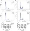
Representative flow cytometric analysis of the cell cycle from the three independent siNEG and 34a-mimic transfections in MDA-MB-231 (a) or Hs578T (b) cells using propidium iodide staining. Percentage of cells within each phase of the cell cycle is indicated. A slight increase in G0/G1 cell-cycle arrest is observed in cells transfected with the miR-34a mimic. The column graph shows the average of all the independent transfections ± SEM performed in triplicate
Fig. 6
Fig. 6
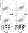
Representative flow cytometric analysis of apoptosis from the three independent siNEG and 34a-mimic transfections in MDA-MB-231 (a) or Hs578T (b) cells using Annexin V and propidium iodide staining. Numbers within the graph represent the percentage of cells detected within a quadrant. No significant change in the percentage of pre-apoptotic (lower right quadrant) or apoptotic (upper right quadrant) cells is observed between the 34a-mimic and siNEG transfections. Split column graph shows the average of all the independent transfections ± SEM performed in triplicate
Fig. 7
Fig. 7
miR-34a and AXL mRNA inversely correlated in basal-like breast cancer patient tumor samples. Scatterplot depicting normalized AXL and miR-34a expression for the 26 basal-like breast cancer patient samples. The inverse relation in expression was determined through a Spearman nonparametric correlation analysis. Open squares indicate those samples that were identified as having p53 mutations

References

    1. Rakha EA, Ellis IO. Triple-negative/basal-like breast cancer: review. Pathology. 2009;41:40–47. - PubMed
    1. Venkitaraman R. Triple-negative/basal-like breast cancer: clinical, pathologic and molecular features. Expert Rev Anti-cancer Ther. 2010;10:199–207. - PubMed
    1. Charafe-Jauffret E, Ginestier C, Monville F, Finetti P, Adélaïde J, Cervera N, Fekairi S, Xerri L, Jacquemier J, Birnbaum D, Bertucci F. Gene expression profiling of breast cancer cell lines identifies potential new basal markers. Oncogene. 2006;25:2273–2284. - PubMed
    1. Kao J, Salari K, Bocanegra M, Choi YL, Girard L, Gandhi J, Kwei KA, Hernandez-Boussard T, Wang P, Gazdar AF, Minna JD, Pollack JR. Molecular profiling of breast cancer cell lines defines relevant tumor models and provides a resource for cancer gene discovery. PLOS One. 2009;4:1–16. - PMC - PubMed
    1. Kreike B, Van Kouwenhove M, Horlings H, Weigelt B, Peterse H, Bartelink H, Van de Vijver MJ. Gene expression profiling and histopathological characterization of triple-negative/ basal-like breast carcinomas. Breast Cancer Res. 2007;9:R65. - PMC - PubMed

MeSH terms