. 2011 Jun;38(6):3039-49.
doi: 10.1118/1.3589138.

The importance of tissue segmentation for dose calculations for kilovoltage radiation therapy

Affiliations

The importance of tissue segmentation for dose calculations for kilovoltage radiation therapy

Magdalena Bazalova et al. Med Phys. 2011 Jun.

Abstract

Purpose: The aim of this work was to evaluate the effect of tissue segmentation on the accuracy of Monte Carlo (MC) dose calculations for kilovoltage radiation therapy, which are commonly used in preclinical radiotherapy studies and are also being revisited as a clinical treatment modality. The feasibility of tissue segmentation routinely done on the basis of differences in tissue mass densities was studied and a new segmentation scheme based on differences in effective atomic numbers was developed.

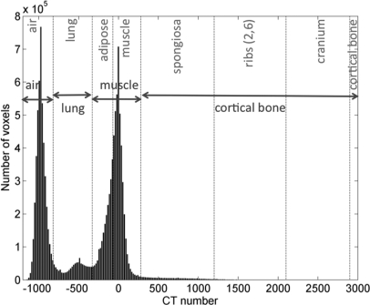
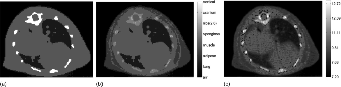
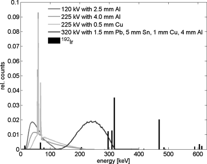
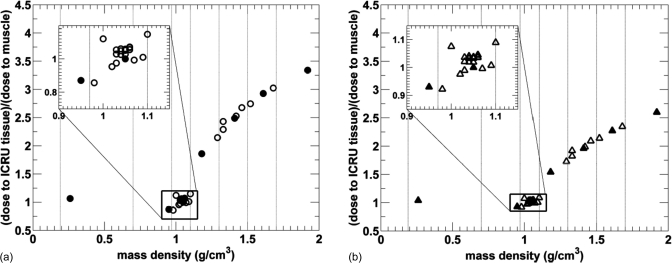
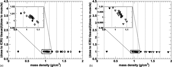
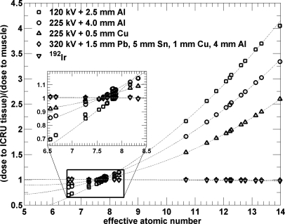
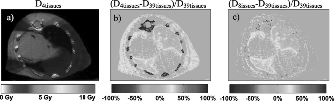
Methods: MC dose calculations in a cylindrical mouse phantom with small cylindrical inhomogeneities consisting of 34 ICRU-44 tissues were performed using the EGSnrc/BEAMnrc and DOSXYZnrc codes. The dose to tissue was calculated for five different kilovoltage beams currently used in small animal radiotherapy: a microCT 120 kV beam, two 225 kV beams filtered with either 4 mm of Al or 0.5 mm of Cu, a heavily filtered 320 kV beam, and a 192Ir beam. The mean doses to the 34 ICRU-44 tissues as a function of tissue mass density and effective atomic number and beam energy were studied. A treatment plan for an orthotopic lung tumor model was created, and the dose distribution was calculated for three tissue segmentation schemes using 4, 8, and 39 tissue bins to assess the significance of the simulation results for kilovoltage radiotherapy.

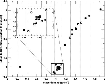
Results: In our model, incorrect assignment of adipose tissue to muscle caused dose calculation differences of 27%, 13%, and 7% for the 120 kV beam and the 225 kV beams filtered with 4 mm Al and 0.5 mm Cu, respectively. For the heavily filtered 320 kV beam and a 192Ir source, potential dose calculation differences due to tissue mis-assignment were below 4%. There was no clear relationship between the dose to tissue and its mass density for x-ray beams generated by tube potentials equal or less than 225 kV. A second order polynomial fit approximated well the absorbed dose to tissue as a function of effective atomic number for these beams. In the mouse study, the 120 kV beam dose to bone was overestimated by 100% and underestimated by 10% for the 4 and 8-tissue segmentation schemes compared to the 39-tissue segmentation scheme, respectively. Dose to adipose tissue was overestimated by 30% and underestimated by 10%, respectively. In general, organ at risk (OAR) doses were overestimated in the 4-tissue and the 8-tissue segmentation schemes compared to the 39-tissue segmentation.

Conclusions: Tissue segmentation was shown to be a key parameter for dose calculations with kilovoltage beams used in small animal radiotherapy when an x-ray tube with a potential < or = 225 kV is used as a source. A new tissue segmentation scheme with 39 tissues based on effective number differences derived from mass density differences has been implemented.


Figures

Figure 1
Figure 1
Dose distribution calculated for five uniformly spaced 5 mm diameter 120 kV beams filtered with 2.5 mm of Al in a 2 cm diameter phantom with a 5 mm cortical bone heterogeneity: the isocenter axial (a), coronal (b), and sagittal slices (c).
Figure 2
Figure 2
Histogram of CT numbers with the corresponding 4-tissue (in blue) and 8-tissue (in red) material assignment of the microCT data set. Dashed lines define air, lung, adipose, muscle, spongiosa, ribs (2,6), cranium, and cortical bone as used in the 8-tissue segmentation model. Adipose and muscle are combined into muscle and spongiosa, ribs (2,6), cranium, and cortical bone are combined into cortical bone in the 4-tissue segmentation model.
Figure 3
Figure 3
Demonstration of tissue segmentation for lower (a) and higher (b) effective atomic number (Z) intervals in small animal dose calculations using 39 tissue types. Z is plotted as a function of mass density for ICRU tissues (squares), for tissue types required to achieve 2% dose calculation accuracy for a 120 kV beam based on Z differences (circles), and for the center of bins for segmentation with 0.02 g∕cm3 mass density bins (open triangles). Tissue types used for dose calculations are shown in closed triangles and separated by solid lines.
Figure 4
Figure 4
Tissue segmentation with 4 tissues (a), 8 tissues (b), and 39 tissues based on effective atomic number differences (c).
Figure 5
Figure 5
Spectral distributions of the photon beams investigated in the study. The 120 kV beam is a circular beam of 5 mm in diameter and the 225 kV beams and the 320 kV beam are (5 × 5) mm rectangular beams. The 192Ir spectrum is the bare spectrum.
Figure 6
Figure 6
Relative dose to tissue as a function of tissue mass density for 34 ICRU tissues from a 120 kV beam with  = 55 keV. Tissues used in 4-tissue and 8-tissue model small animal dose calculations are plotted with filled symbols and vertical lines indicated.
Figure 7
Figure 7
Relative dose to tissue as a function of tissue mass density for 34 ICRU tissues from a 225 kV beam filtered by 4 mm of Al with  = 77 keV (a) and 0.5 mm of Cu with  = 92 keV (b). Tissues used in 4-tissue and 8-tissue model small animal dose calculations are plotted with filled symbols and vertical lines indicated the tissue segmentation ranges. Statistical uncertainties of <1.4% are not plotted for clarity.
Figure 8
Figure 8
Relative dose to tissue as a function of tissue mass density for 34 ICRU tissues from irradiation with an extensively filtered 320 kV beam with  = 237 keV (a) and a 192Ir source with <E> = 346 keV (b). Tissues used in 4-tissue and 8-tissue model small animal dose calculations are plotted with filled symbols and vertical lines indicated the tissue segmentation ranges. Statistical uncertainties of <1.4% are not plotted for clarity.
Figure 9
Figure 9
Relative dose to tissue as a function of tissue effective atomic number for 34 ICRU tissues from irradiation with photon beams used for conformal small animal radiation therapy. The quadratic fits are plotted with dashed lines.
Figure 10
Figure 10
Monte Carlo dose distribution for an orthotopic lung tumor with the standard 4-tissue segmentation scheme (a) for a 120 kVp plan. The percentage difference maps (D4tissuesD39tissues)∕D39tissues and (D8tissuesD39tissues)∕D39tissues are shown in (b) and (c), respectively. The PTV is delineated in red.
Figure 11
Figure 11
DVH for the PTV, the spinal cord, the heart and the left and the right lung for 120 kVp dose calculations with the 39-tissue segmentation scheme (solid curves), with the 4-tissue segmentation scheme (dashed curves), and with the 8-tissue segmentation scheme (dotted curves).

References

    1. Hillman G. G., Maughan R. L., Grignon D. J., Yudelev M., Rubio J.,Tekyi-Mensah S., Layer A., Che M. X., and Forman J. D., “Neutron or photon irradiation for prostate tumors: Enhancement of cytokine therapy in a metastatic tumor model,” Clin. Cancer Res. 7, 136–144 (2001). - PubMed
    1. Khan M., Hill R., and Vandyk J., “Partial volume rat lung irradiation: An evaluation of early DNA damage,” Int. J. Radiat. Oncol. Biol. Phys. 40, 467–476 (1998).10.1016/S0360-3016(97)00736-0 - DOI - PubMed
    1. Stojadinovic S., Low D. A., Vicic M., Mutic S., Deasy J. O., Hope A. J., Parikh P. J., and Grigsby P. W., “Progress toward a microradiation therapy small animal conformal irradiator,” Med. Phys. 33, 3834–3845 (2006).10.1118/1.2349693 - DOI - PubMed
    1. Wong J., Armour E., Kazanzides P., Iordachita U., Tryggestad E., Deng H., Matinfar M., Kennedy C., Liu Z. J., Chan T., Gray O., Verhaegen F., McNutt T., Ford E., and DeWeese T. L., “High-resolution, small animal radiation research platform with x-ray tomographic guidance capabilities,” Int. J.Radiat. Oncol. Biol. Phys. 71, 1591–1599 (2008).10.1016/j.ijrobp.2008.04.025 - DOI - PMC - PubMed
    1. Matinfar M., Ford E., Iordachita I., Wong J., and Kazanzides P., “Image-guided small animal radiation research platform: Calibration of treatment beam alignment,” Phys. Med. Biol. 54, 891–905 (2009).10.1088/0031-9155/54/4/005 - DOI - PMC - PubMed

Substances